 |
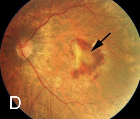
| A macular hemorrhage and fibrovascular membrane (arrow) in a 67-year-old -10.50D myopic woman. Photo: Julie Poteet, OD. Click image to enlarge. |
A recent population-based analysis provides additional insights on the prevalence and causes of visual acuity and visual field loss in highly myopic eyes. Participants of the Beijing Eye Study who underwent an ophthalmological and systemic examination that included frequency doubling technology perimetry (FDT) were involved in this research (n=4,439).
Among the 8,878 eyes in this study, 79 (0.9%) eyes had prior cataract surgery. Thirteen of these eyes underwent ocular biometry in 2022, and none of them had an axial length of more than 24.0 mm. The remaining 66 eyes were excluded from the current study since they had not undergone biometry and, as a result, did not have reliable data regarding the presence and degree of pre-operative axial myopia, according to the study authors.
Out of the total study population, 2,695 (60.7%) were reexamined 10 years later and biometry was used to measure the axial length in their right eyes. Researchers defined high myopia as a refractive error of ≤-6D or axial length >26.0mm. The main outcome measure was the prevalence of vision impairment causes.
The current analysis included 212 highly myopic eyes (154 participants). The mean refractive error was -9.87D ± 3.70D (median: -8.63D; range: -20.87D to -6.00D) and mean axial length (if measured in 2011) was 27.2mm ± 1.3mm (median: 26.5mm; range: 26.01mm to 30.88mm).
Study authors reported that moderate/severe vision impairment (MSVI)—defined as best corrected visual acuity (BCVA) from <6/18 to 3/60—was present in forty eyes (18.9%). Blindness (BCVA <3/60 or <10° visual field around central fixation) was observed in 10 eyes (4.7%).
Among these highly myopic patients, data showed that BCVA decreased with more myopic refractive error. Myopic macular degeneration (MMD), defined as stage 3 or 4, and optic neuropathy were the most common causes of MSVI and blindness, according to the research team that reported that MMD prevalence increased from 1.6% in the refractive error group of -6.00 to ≥-7.00D to 64.0% in the group of <-15.0D. Additionally, they found that it correlated with a “higher prevalence of concomitant optic neuropathy of both, glaucomatous and non-glaucomatous type.”
Optic neuropathy of a glaucomatous (glaucoma-like) and non-glaucomatous type contribute to the loss in visual acuity and visual field in highly myopic eyes, the researchers reported in their recent paper. “Their prevalence of optic neuropathy non-linearly increases with more marked myopia refractive error and reached 36% in eyes with a myopic refractive error of more than -15 diopters,” they noted.
“The pathogenesis of high myopia associated optic neuropathies may be connected with axial elongation-related changes in the lamina cribrosa and parapapillary region and with an axial elongation-associated increase in the distance between the retinal ganglion cells and the optic disc.”
Jonas JB, Jonas RA, Xu J, et al. Prevalence and Cause of Loss of Visual Acuity and Visual Field in Highly Myopic Eyes. The Beijing Eye Study. Ophthalmology. September 4, 2023 [Epub ahead of print]. |
