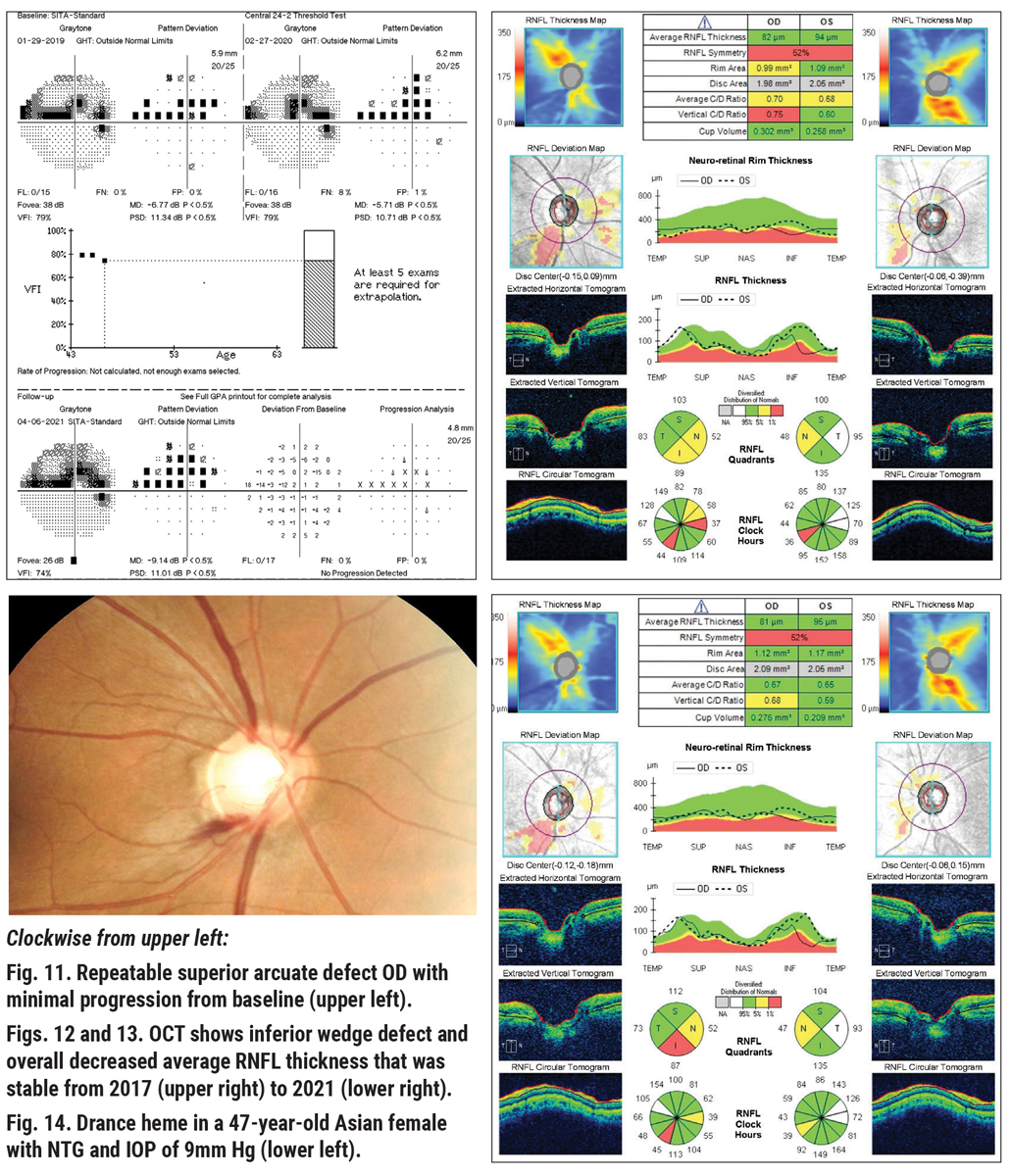
 |
| Disc hemorrhages often appears at or near glaucomatous disc/retina sites of change, RNFL defect border and site of peripapillary atrophy, suggesting its development is closely related to structural damage of the disc and/or retina. Photo: Sarah B. Klein, OD. Click image to enlarge. |
Early detection of glaucoma is crucial to prevent or delay irreversible visual impairment. Some studies on primates have indicated disc and lamina cribrosa changes to occur before deterioration happens in visual function and changes happen within the retinal nerve fiber layer, with some lag between both events. Similarly, researchers of a recent study wanted to identify factors associated with structural and functional deterioration progression, since such changes may go unnoticed in open-angle glaucoma (OAG) patients with lower than normal intraocular pressure (IOP).
Specifically, factors associated with disc/retina deterioration in stereo fundus images before that of the visual field were observed. The prospective study included 90 eyes of 90 OAG patients and baseline IOPs of less than 15mm Hg. The five-year study was conducted without treatment of participants. IOP measurements were collected every three months and fundus photographs were collected every six.
Average age of patients was 54 years. During the five-year follow-up, “field first” deterioration probability was determined to be 49±6.6%, while “structure first” manifestation was 33±6.4%. Prior to deterioration, the presence of a disc hemorrhage was associated with the structure first subtype, while older age was linked with the field first group.
The authors note that their study is unique in its absence of IOP-lowering therapy before more deterioration of the disc/retina or visual field was confirmed. The only other study that has been conducted without treatment was the Ocular Hypertension Treatment Study, which found 89 out of 819 untreated eyes to develop glaucoma. Of those, 57% displayed disc/retina deterioration first, 33% had visual field deterioration first and the remaining patients had concurrent deterioration of both. This contrasts with the current study’s lower percentage of structure first deterioration.
Glaucomatous eyes more frequently present with disc hemorrhages than healthy ones, but the mechanism for its development is not yet understood. One possibility is that vascular abnormalities may play a role, as suggested by hemorrhage frequency being greater in those diabetes, hypertension and lower systolic blood pressure. Aspirin use and antiplatelet drugs are also reportedly linked with hemorrhage, thus raising the possibility of a link between abnormal microcirculatory status that may cause a predisposition for microvasculature disruption of tissue around the optic disc. However, the participants in this study did not have circulatory abnormalities, so disc hemorrhage was unlikely to occur first, triggering subsequent events.
In their paper for the British journal Eye, the authors offer clinical advice based on their results: “Although both visual filed and disc/retina must be vigilantly monitored to accurately track the disease’s progression, our results would suggest that when disc hemorrhage is observed in OAG patients with lower normal IOP, more attention should be paid to disc/retina findings for early detection of progression, while in older patients without hemorrhage attention should be paid to visual field.”
Sakata R, Araie M, Yoshitomi T; for lower normal pressure glaucoma study members in Japan Glaucoma Society. Factors associated with visual field or structure progression occurring first in a prospective study on patients with untreated open-angle glaucoma with normal intraocular pressure. Eye (Lond). October 5, 2023. [Epub ahead of print]. |
