 |
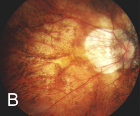
| In eyes with pathologic myopia, MNV-related atrophy may develop and progress differently based on the atrophic pattern. Photo: Julie Poteet, OD. Click image to enlarge. |
Between 5.2% and 11.3% of individuals with pathologic myopia have myopic macular neovascularization (MNV), a vision-threatening lesion that will inevitably result in atrophy as the condition progresses. A recent small study identified three patterns of MNV-related atrophy in this patient population, described as multiple-atrophic, single-atrophic and exudation-related, each of which had a unique course of progression.
The study included 27 eyes of 26 patients (23 female) with MNV who were followed for an average of 78 months from onset to progression to macular atrophy. The mean age of patients was 67 years, the mean axial length was 29mm and all patients were treated with anti-VEGF injections. The researchers reviewed a longitudinal series of autofluorescence and OCT images to examine the pattern of MNV-related atrophy in each eye. Change in best-corrected visual acuity (BCVA) was also determined for each atrophic pattern.
Multiple-atrophic was the most common MNV-related atrophy pattern observed among the cohort, affecting 17 eyes (63%) and presenting as small atrophies occurring at multiple sites around the MNV edge. An equal number of eyes had a single-atrophic or exudation-related atrophy pattern (five eyes [18.5%] for each). Those with the single-atrophic pattern presented with atrophies occurring on only one side of the MNV edge, and the exudation-related atrophy pattern was characterized by atrophy occurring within a previous serous exudation or hemorrhagic area and slightly away from the MNV edge.
The researchers noted that, overall, mean BCVA at three years was significantly poorer than baseline, although there were distinctions between average BCVA changes for each of the three atrophic patterns. “Eyes with atrophies in multiple-atrophic and exudation-related patterns progressed to large macular atrophies involving the central fovea and showed a decrease of BCVA during the three-year follow-up period,” the study authors wrote in their paper, published in Retina. On the other hand, they reported that “eyes with single-atrophic pattern had a sparing of the fovea and had good recovery of the BCVA.”
The authors suggest that, due to differences in progression between the different atrophic patterns, specific treatment protocols may be necessary for each type. They offered the following clinical guidance in their paper: “For multiple-atrophic pattern, routine fundus autofluorescence examinations are recommended to monitor the progression of atrophy and change in the BCVA. For single-atrophic pattern, patients should be strongly recommended to return for examinations when they notice any change in vision due to a recurrence. For exudation-related pattern, more attention should be paid to decreasing the exudative changes urgently.”
Xie S, Lu H, Chen C, et al. Morphological characteristics and progression patterns of macular neovascularization-related atrophy in eyes with pathologic myopia. Retina. June 19, 2023. [Epub ahead of print]. |