 |
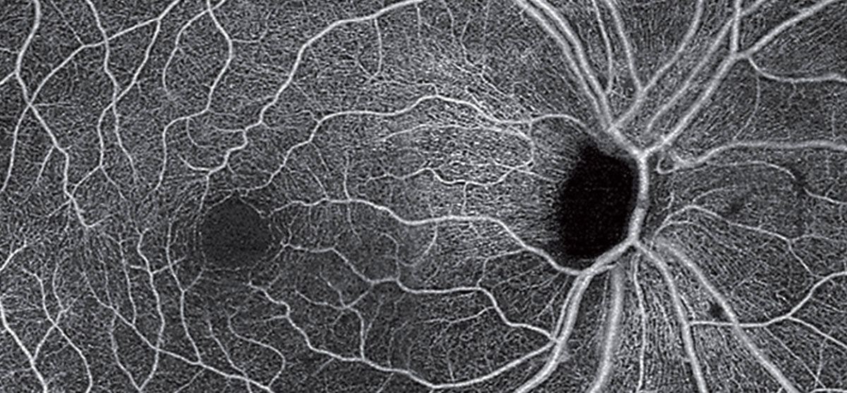
| OCT-A montage of an unrelated patient (with POAG) showing loss of radial peripapillary capillaries and the superficial capillary plexus. Photo: Carolyn Majcher, OD. Click image to enlarge. |
As researchers continue to uncover ocular manifestations of central nervous system diseases, it’s important for eye doctors to understand and recognize these connections. Parkinson’s disease patients present with reduction of the neuroretina, but there are few studies on the tissue’s integrity in essential tremor patients. Current diagnostic criteria for the two conditions are clinical; the lack of disease-specific diagnostic markers for both processes may lead to delayed diagnosis and even misdiagnosis. Researchers in Spain recently evaluated the retinal vascular plexus among these patients with swept-source optical coherence tomography (SS-OCT).
The study consisted of a total of 42 Parkinson’s and 26 essential tremor patients and 146 controls who all underwent retinal evaluation using SS-OCT plus OCT angiography (OCT-A). The researchers assessed the macular and peripapillary retinal nerve fiber layer (RNFL) and ganglion cell layer (GCL) as well as macular vasculature. They performed logistic regression analysis to ascertain whether any of the structural parameters evaluated in the study had the ability to differentiate between healthy eyes and those with essential tremor or Parkinson’s disease.
Parkinson’s patients presented a reduction in macular RNFL, macular GCL and peripapillary RNFL vs. healthy controls and in macular RNFL and peripapillary RNFL vs. essential tremor patients. Essential tremor patients showed a significant reduction in macular GCL vs. controls. The researchers found no differences in the macular vasculature between groups. Retinal hypoperfusion was not observed, and the researchers believe that it is unlikely that it plays a role in the retinal degeneration of these diseases. Predictive diagnostic variables were significant only for Parkinson’s disease, and a linear discriminant was obtained with an area under the ROC curve of 0.796.
The researchers noted that their findings about use of OCT-A “could be useful as a supplementary tool in diagnosing Parkinson’s disease. However, our regression analysis did not yield any results for essential tremor patients, making the diagnostic potential of OCT in patients with early-stage suspected tremor (essential tremor vs. early Parkinson’s) uncertain at this time.”
While its utility in the diagnosis of overlapping essential tremor and Parkinson’s disease is still uncertain, SS-OCT analysis of retinal measurements in these conditions has proven to be a technique that is fast, noninvasive and cost-effective.
Satue M, Castro L, Vilades E, et al. Ability of swept-source OCT and OCT-angiography to detect neuroretinal and vasculature changes in patients with Parkinson disease and essential tremor. Eye (Lond). June 1, 2022. [Epub ahead of print]. |
