 |
This study found a much higher prevalence rate of imaging artifacts in high myopes vs. non-myopes, which researchers suggest may be due to various factors such as globe elongation and posterior pole disruption. The above abnormal OCT shows poor segmentation in both the heat maps and tomograms in a high myope. Photo: Andrew Rouse, OD. Click image to enlarge. |
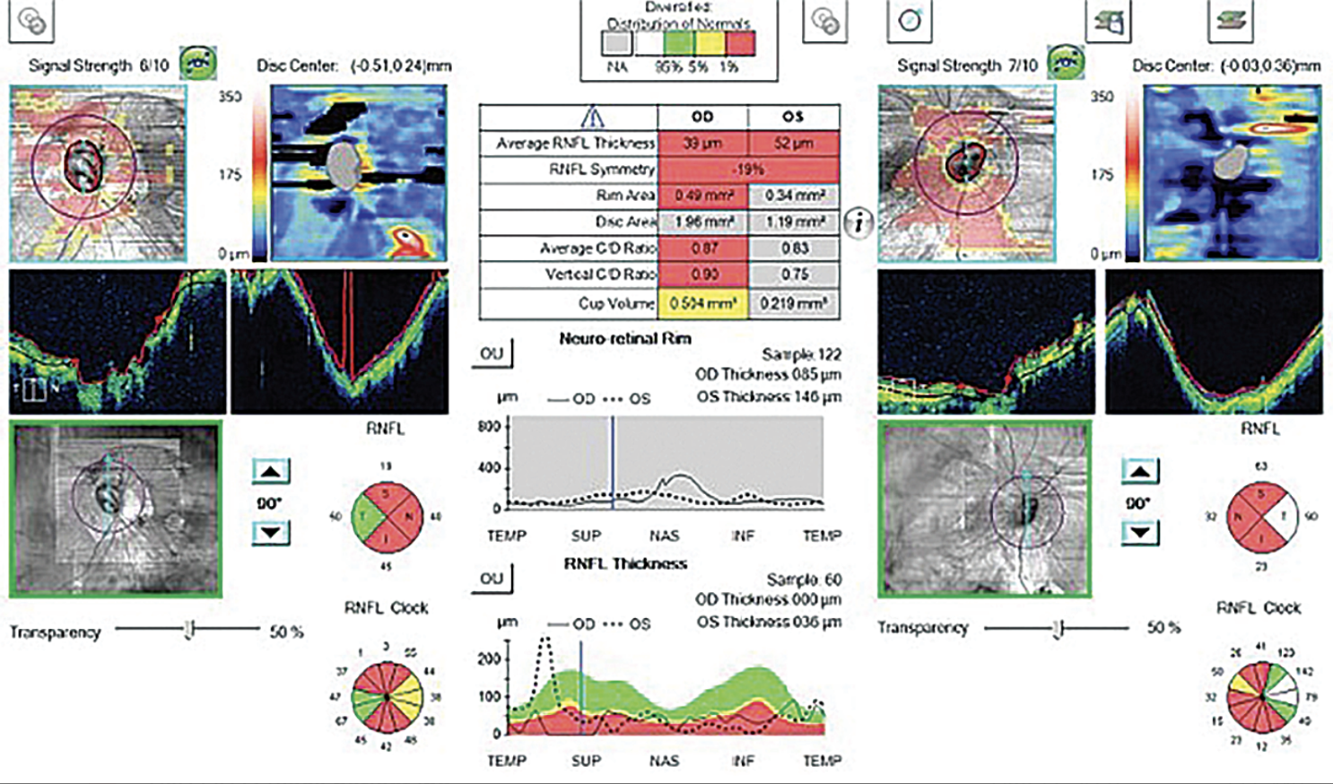
While OCT is a useful and often reliable tool for diagnosing and monitoring glaucoma, it’s important that clinicians recognize the presence of imaging artifacts to avoid inaccurate scan interpretation. A new study in the Journal of Glaucoma suggests this may be especially crucial when assessing glaucoma patients who also have high myopia, as it noted a higher prevalence of imaging artifacts among these patients compared with non-myopes.
The prospective study enrolled 226 patients with glaucoma and/or high myopia, who were divided into four groups based on their refractive status and which condition(s) they had: non-high myope controls, non-high myope glaucoma patients, highly myopic patients and highly myopic patients with glaucoma. For each patient, peripapillary retinal nerve fiber layer (RNFL) scan images were inspected for the presence of artifacts.
The prevalence of OCT artifacts among the cohort was significantly higher in high myopes (51.9%) than in non-high myopes (18.6%). However, the most common type of artifact differed between the two; in high myopes, retinal pathology-related artifact was the most common type (23%; 4% in non-high myopes), and in non-myopes, the most common was outer RNFL border misidentification (11%; 19.4% in high myopes).
The researchers also determined three prominent predictive factors for the presence of OCT artifacts: a longer axial length (odds ratio [OR]: 1.82), a higher pattern standard deviation (OR: 1.19) and thinner RNFL (OR: 0.95).
“Overall, OCT artifact is relatively uncommon in a non–highly myopic normal population (4.9%),” the researchers wrote in their study. “However, the prevalence of OCT artifacts becomes much higher with a patient having glaucoma (33.3%), high myopia (43.5%) or both (63.0%).”
They go on to explain a few common reasons why artifacts are more common in these patients. For example, regarding outer RNFL border misidentification in high myopes, the study authors note that “extreme elongation of the globe may result in an inadequately captured image due to exceeding the diopter compensation of the device or from insufficiently reflected image signals.” They add, “Distortion of the posterior pole may also result in a highly curved peripapillary retinal image on OCT and could hinder the segmentation algorithm from identifying the outer border of the RNFL accurately.” Furthermore, the authors note that in high myopes, irregularity in the contour of the peripapillary region can lead to a variable focusing effect and irregular light scattering, increasing the likelihood of segmentation errors.
Retinal pathology-related artifact, the most common observed in highly myopic patients in this study, may appear as the loss or disruption of the retinal pigment epithelium layer, RNFL plaques, retinal thinning or abnormal retinal sloping.
Imaging acquisition artifact was among the less common types observed and can often be reduced through more technician training and experience, the researchers noted. However, they did find that motion artifact had a prevalence of 9.3% among high myopes vs. 0.8% in non-myopes, which they hypothesized “could reflect the difficulties in reaching an adequate focal plane, needing frequent adjustments to obtain an OCT image in high myopes as their eyeballs are more elongated than a typical normal eye.”
In light of these findings, the researchers advise that special care should be taken when interpreting OCT scans of patients with glaucoma, high myopia or both to ensure accurate assessment and informed clinical decision-making.
Poon LYC, Wang CH, Lin PW, et al. The prevalence of optical coherence tomography artifacts in high myopia and its influence on glaucoma diagnosis. J of Glaucoma. 2023;32:7. |
