 |
|
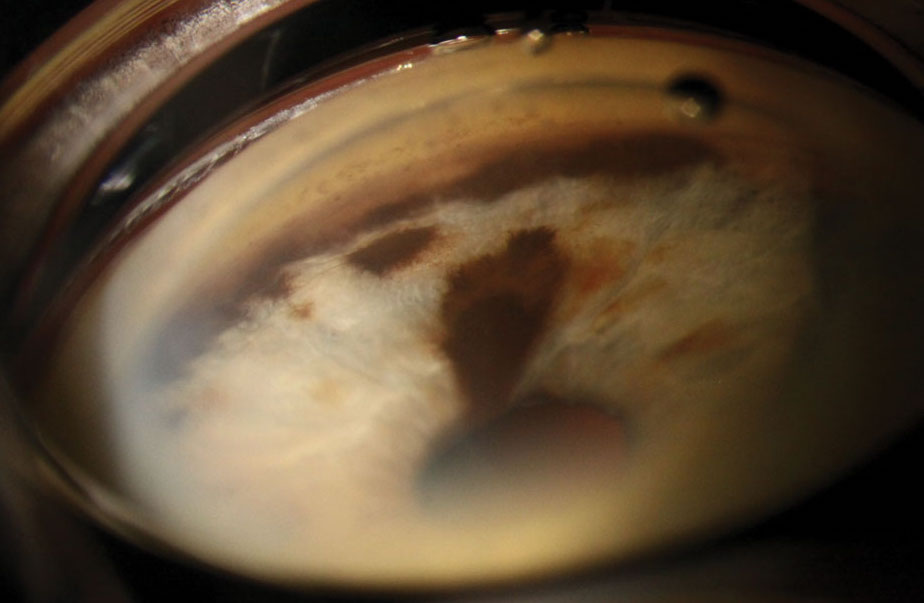
Compared to eyes with an iris nevus, this study found that those with iris melanoma had elevated IOPs, higher cup-to-disc ratios and were more likely to have a secondary glaucoma diagnosis at presentation. Photo: Christine Sindt, OD. Click image to enlarge. |
Multiple studies and case reports have identified clinical features besides tumor growth that can help eyecare providers differentiate an iris melanoma from an iris nevus, two of which include elevated intraocular pressure (IOP) and cup-to-disc (c/d) ratio. Given that iris melanoma is known to interfere with the trabecular meshwork and increase risk of secondary glaucoma, researchers recently wanted to investigate how these two parameters compare between eyes with the malignant condition and those with an iris nevus, as well as whether measurements were notably greater in affected eyes.
This retrospective study spanned almost a decade and included patients treated for iris melanoma and iris nevus at UCLA's Stein Eye Institute. Researchers compared IOP, c/d ratio and glaucoma diagnosis between affected and unaffected eyes among 39 subjects with iris melanoma and 40 with an iris nevus.
The results indicated significantly higher IOP and c/d ratio in affected eyes of patients with iris melanoma compared to their unaffected counterparts. On average, melanoma-affected eyes displayed an IOP of 18.8mm Hg vs. 14.6mm Hg in nevus-affected eyes. The average c/d ratio in eyes with iris melanoma was 0.36 compared to 0.24 in eyes with iris nevus.
The study also identified differences in IOP and c/d ratio between affected and unaffected eyes in patients with iris melanoma. The average IOP of the unaffected eye in these patients was 16.3mm Hg, more than 2mm Hg less than the affected eye. The average c/d ratio of the unaffected eye in iris melanoma was also slightly less than the affected eye at 0.25. Conversely, in eyes with an iris nevus, no differences were noted between the average IOP or c/d between affected and unaffected eyes.
Another notable but perhaps unsurprising finding is that nearly a third of patients with iris melanoma had a secondary glaucoma diagnosis at the time of presentation. All these patients were on IOP-lowering medication prior to their initial lesion evaluation. Even in patients without secondary glaucoma, the researchers reported greater IOP in eyes with iris melanoma than in those with an iris nevus.
In their paper on the study, published in Clinical Ophthalmology, the authors reported that the area under the receiver operating characteristic (ROC) curves—a measure of the predictive ability of one a given variable—for absolute IOP values, IOP differences and c/d ratio differences “were all significant” to help distinguish iris melanoma from iris nevus, “with the ROC curve for absolute IOP demonstrating the best diagnostic utility.”
While it was already known that patients with iris melanoma may present with elevated IOP, these findings highlight that increasing IOP asymmetry—and, to a lesser extent, CDR asymmetry—between both eyes may be a helpful indicator of the malignant condition.
Asymmetry in pressure and cup-to-disc ratio “are two easily identifiable parameters in the clinical evaluation that may indicate a high likelihood of the diagnosis of iris melanoma,” the authors concluded. They surmised that this research may help guide future studies that could lead to standardized diagnostic protocols involving these biometric parameters.
| Click here for journal source. |
Kong AW, Au A, Song W, Oh AJ, McCannel TA. Intraocular pressure and cup-to-disc ratio asymmetry in diagnosing iris melanoma. Clin Ophthalmol. October 15, 2024. [Epub ahead of print]. |

