 |
|
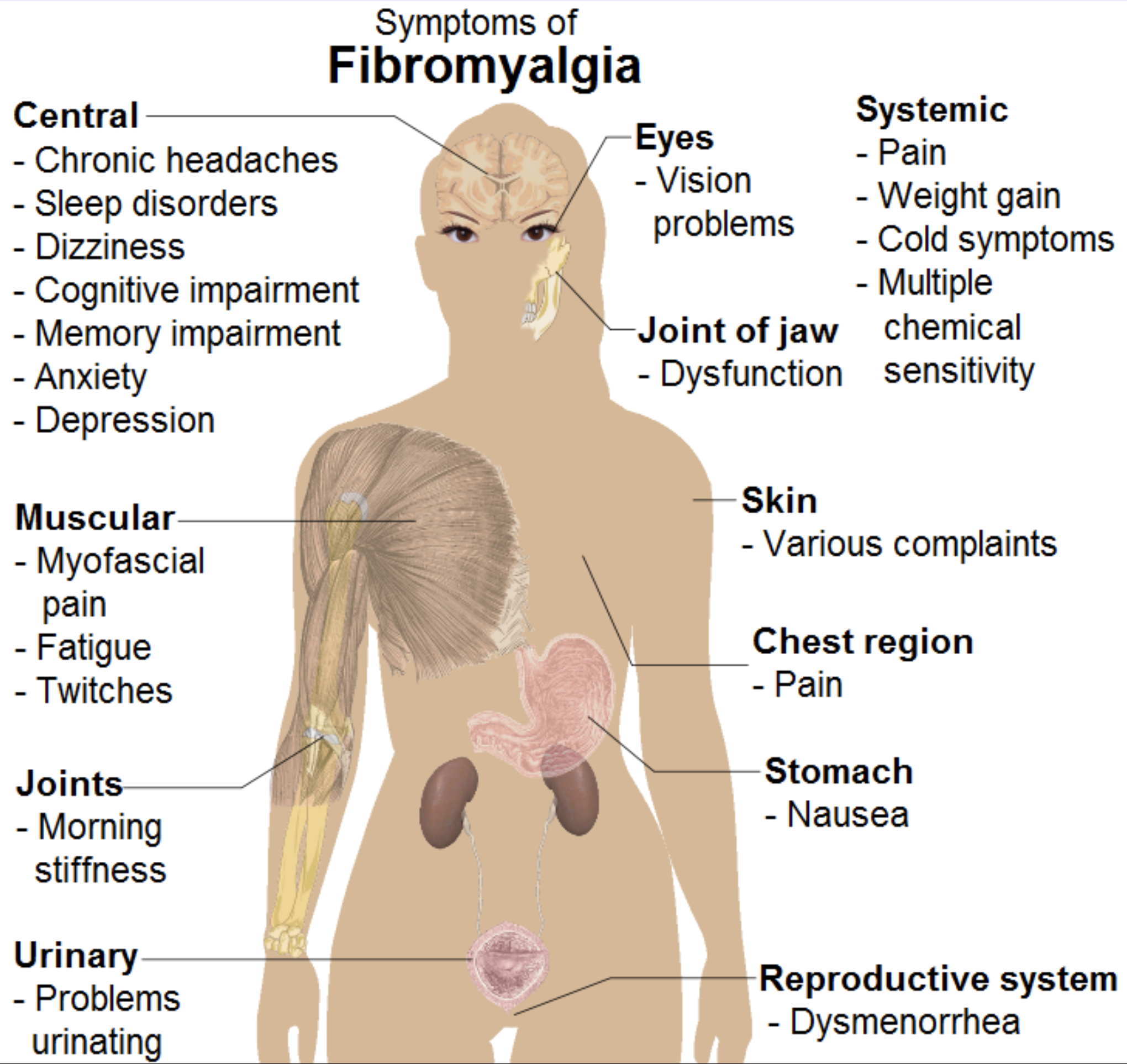
Spectralis OCT was able to effectively differentiate between healthy patients and those with FM. Photo: Wikimedia Commons. Click image to enlarge. |
Editor’s Note: As part of our “Year in Review” retrospective, we’ve selected the top 30 news stories of the year and are re-sharing them as we close out 2022. Follow along as we count down to number 1!
This story was originally published on September 1, 2022.
No. 5 biggest news story of 2022:
The pathophysiology of fibromyalgia isn’t fully understood, but digital imaging of the neuroretina may shed some light on the condition. Researchers recently found OCT-observable retinal changes in FM patients; in a separate study, the same group then calculated two different linear discriminant functions (LDFs)—a way to measure variance—to improve the specificity of OCT retinal parameters in FM diagnosis.
The study included 50 patients with FM and 232 healthy controls. All participants underwent retinal evaluation using swept-source OCT angiography (Triton Plus) and spectral-domain OCT (Spectralis). They reported that the Spectralis was able to differentiate between controls and FM patients. The researchers observed no significant differences in the macular vascular plexus between FM patients and controls, but they did note that the vascular density in the superior sector showed strong inverse correlation with disease duration.
With Triton OCT, FM patients demonstrated significant peripapillary RNFL thinning in the temporal sector. With Spectralis OCT, patients demonstrated significantly decreased peripapillary RNFL in the superonasal, nasal, inferonasal, temporal and inferotemporal sectors. The researchers noted in their paper that the LDF calculated for the Spectralis showed an area under the curve (a measure of predictive accuracy) of 0.968.
They concluded that FM patients present with observable RNFL thinning on swept-source and spectral-domain OCT, but they also demonstrate a macular vascular density similar to that of healthy controls. The researchers wrote in their paper that this suggests “retinal structural changes aren’t due to retinal hypoperfusion” and supports the hypothesis that neurodegeneration is present in FM.
The group also noted in their paper that, based on the Spectralis’ AUROC, “The LDF that combines several RNFL parameters obtained with Spectralis OCT gives this device a powerful ability to differentiate between healthy individuals and individuals with FM.”
Garcia-Martin E, Tello A, Vilades E, et al. Diagnostic ability and capacity of optical coherence tomography-angiography to detect retinal and vascular changes in patients with fibromyalgia. J Ophthalmol. 2022;3946017:1-8. |
