 |
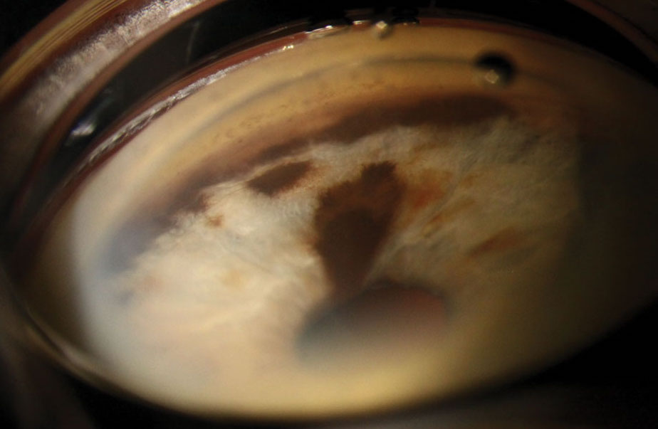
| Iris melanoma can have devastating visual outcomes if it's not detected soon enough for aggressive intervention. Photo: Christine Sindt, OD. Click image to enlarge. |
A rare form of intraocular malignancy, iris melanoma, accounts for just 4% of the roughly 7,000 cases of uveal melanomas diagnosed globally each year. Most affected patients live in Europe and North America, which are shown to be high ultraviolet environments, and 90% of cases originate in the choroid. Because research on the disease is lacking, authors of a recent study observed data from a group of patients with iris melanocytic tumors to identify demographics, diagnostic features, management and outcomes. Their findings emphasized the importance of making eye care accessible on a global level.
Records from a 20-year period (1999-2018) were collected from a clinic in Auckland, New Zealand. The 51 patients included in the study were diagnosed with or suspected to have iris melanoma, and 98% were European and from New Zealand. The following data were analyzed: demographics, tumor characteristics, histology, genetics, treatment modalities, recurrence, metastasis, five-year and overall survival.
The median age at presentation was 58 years, and tumors involved a median of two clock hours of the iris. In 45.8% of patients, the posterior tumor margin extended to the anterior chamber angle, and half of the patients had tumors with no distinctive shape that were classified as “geographic.” The most frequent clinical features associated with the tumors were ectropion uvea (41.2%), corectopia (39.2%), cataract (37.3%) and secondary glaucoma (23.5%).
Patients were most commonly managed initially with observation (54.9%) and then with iridectomy/excision biopsy (29.4%), irido-cyclectomy (7.8%), plaque radiotherapy (7.8%) and proton beam radiotherapy (7.8%). For 17.6% of patients, enucleation of the eye was eventually needed. Of the 37% of patients who had histology reports, 84% were confirmed to have melanoma. The mean follow-up was 4.2 years, and the median visual acuity after two years post-intervention was 6/7.5.
Current estimations show that approximately 50% of patients with uveal melanoma will eventually die of metastatic disease. The five-year melanoma-related mortality rate of this study cohort was 4%, occurring in two of the 51 patients. The researchers noted that “the proportion of high-grade tumors (T2-4) in this study (4.1%) was significantly lower than that in the United States (25%). This suggests that the free-to-access public hospital ophthalmology service and widespread community optometry services are capable of recognizing iris tumors early to allow patients access to high-quality care promptly.”
The researchers also pointed out that in their study, “there was a high prevalence of more advanced clinical features including ectropion uvea, corectopia, cataract and fewer spindle cell tumors than comparable studies,” which they suggest “may reflect the high ultraviolet exposure in New Zealand and a predominantly white Caucasian population.”
Recognizing the signs of melanocytic lesions and intervening with treatment as early as possible is critical in obtaining better outcomes and reducing the number of patients who lose their eyes to enucleation. The researchers concluded that for European and North American populations, better access to care is needed to promote earlier detection of ocular melanomas such as iris melanoma.
Rapata MEJ, Zhang J, Cunningham WJ, et al. Iris melanocytic tumors in New Zealand/Aotearoa: presentation, management and outcome in a high UV exposure environment. Eye (Lond). March 25, 2022. [Epub ahead of print]. |
