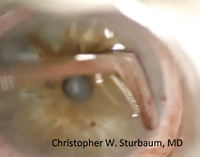
Band keratopathy, a relatively common corneal dystrophy caused by calcification of the anterior stroma and Bowman’s layer, usually occurs between the palpebral fissures, starting peripherally and then progressing to the center. The cornea has rough, white, plaque-like patches separated from the limbus by clear cornea. There may be “holes” in the calcium deposits, which can give it a Swiss cheese appearance.1

|
 |
Symptomatology can vary widely, from asymptomatic patient histories to those who describe decreased vision and severe foreign body sensation (directly proportional to the density of the deposits). There are several possible etiologies; the most common include idiopathic causes, dry eye, chronic uveitis, glaucoma, corneal edema and hypercalcemia. Additionally, conditions to consider in the differential include gout, in-ter-stitial keratitis, primary and secondary calcareous corneal degeneration, calciphylaxis and spheroidal degeneration. Also, consider the potential for an ocular and systemic hypersensitivity reaction characterized by calcium deposition in response to specific antigens or agents.

|
|
|
Click
here for a video of a chelation procedure.
|
Collaboration is Key
Communication with the PCP is essential in managing these patients, as a work-up for hyperparathyroid, sarcoid, gout or renal failure may be needed.2,3 Prolonged exposure to chemicals such as mercury and intraocular substances (e.g., silicone oil) can also cause band keratopathy. These are important to remember—pilocarpine contains mercurial preservatives, for instance, and silicone oil is commonly used in retinal detachment surgery. Thus, it is essential to monitor for corneal changes over time.4
If the patient only experiences mild foreign body sensation with no effect on vision, treating with topical lubrication—as you would treat ocular surface disease—may suffice. As the patient progresses to visual acuity loss, increased foreign body sensation or increased cosmetic concerns, further intervention is warranted. The recommended course is chelation by disodium ethylenediaminetetraacetic acid (EDTA).
In the chelation procedure, the cornea is prepped by gentle epithelial debridement with a spud or scalpel, then a corneal well is placed on the eye and disodium EDTA 3% is added to it. As seen in the accompanying video, the solution alone will dissolve the calcium precipitates, but gentle rubbing of the plaques increases the break-up time. Once completed, the EDTA is rinsed off the cornea and a bandage contact lens is inserted for comfort until the cornea is re-epithelialized.3
Post-op medications include topical antibiotics, NSAIDs and steroids for comfort and treatment of inflammation and corneal edema. These can be stopped once the epithelium is healed and the bandage lens is removed. Additional treatment options include phototherapeutic keratectomy or superficial debridement, which generally restore vision and comfort for most patients with band keratopathy.
In our practice, we had traditionally waited until patient symptoms were severe before EDTA treatment was recommended. Now, we have found that early intervention improves patient comfort and vision, and prevents further stromal scarring. Due to the minimally invasive nature of the treatment, early intervention is often very much appreciated by the patient.
It is important to remember that the recurrence rate of band keratopathy is high. However, the chances can be reduced if the underlying cause is addressed promptly upon diagnosis.
Drs. Claypool and Sturnbaum practice together at Empire Eye in Spokane, Wash.
1. Jhanji V, Rapuano CJ, Vajpayee RB. Corneal calcific band keratopathy. Curr Opin Ophthalmol. Jul 2011;22(4):283-9.
2. Porter R, Crombie AL. Corneal and conjunctival calcification in chronic renal failure. Br J Ophthalmol. May 1973;57(5):339-43.
3. Ehlers J, Shah C. Band Keratopathy. Wills Eye Manual. 2008; 60-62.
4. Available at: http://dro.hs.columbia.edu/bandk.htm.

