Corneal research presented at ARVO has always been a fruitful area of innovation, and this year was no exception––with numerous studies aimed to improve our understanding of graft surgery outcomes, dry eye management and successful lens wear strategies. Highlights are summarized here.
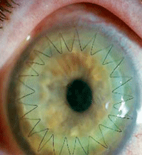
Surgeons who performed more than 15 lamellar procedures per year achieved a markedly better rate of graft survival than those who performed fewer surgeries. Photo: Maynard L. Pohl, OD
(For additional coverage and commentary that focuses on ways to limit corneal morbidity due to inflammation and infection, be sure to look for this month’s Review of Cornea & Contact Lenses.)
Wait a Second, Femtosecond!
One of the most tantalizing, presumed benefits of femto-assisted cataract surgery is the potential safety gains to be had from the reduction in phaco power used––as the laser handles lens fragmentation and reduces dependence on phaco. Is it borne out in practice? A small study suggests not.1649/D0284
In a review of 15 eyes that underwent cataract surgery with or without femto assistance––all performed by the same surgeon and phaco machine––endothelial cell density at one month post-op was only marginally lower in the phaco group (2,696/mm2 ±233/mm2) than in the femto group (2,833/mm2 ±140/mm2). Peripheral endothelial cell loss was significant in both the phaco group (4%, p=0.004) and the femto group (2%, p=0.005).
The authors concluded that femto-assisted cataract surgery does not significantly differ from traditional phaco in early corneal peripheral endothelial cell loss.
Corneal Transplant Sx Update
Recent advances in lamellar corneal surgery have been a boon to corneal graft patients, but there appears to be a learning curve for surgeons, which ultimately impacts outcomes. In a review of more than 23,000 corneal graft procedures spanning 25 years, a total of 2,983 lamellar grafts were identified: 42% endokeratoplasties (posterior corneal endothelial cell grafts), 39% traditional lamellar keratoplasties and 19% deep anterior lamellar keratoplasties (DALKs).1753
Kaplan-Meier graft survival at one year was 74% for endokeratoplasties, 80% for traditional lamellar procedures and 93% for DALKs. Surgeons who performed more than 15 lamellar procedures per year achieved significantly better graft survival than those who performed fewer grafts (p=0.02). A best-corrected acuity of 20/40 or better was achieved in 18% of endokeratoplasties, 34% of traditional lamellar grafts and in 37% of DALKs. The authors concluded that graft survival and visual acuity outcomes are better for penetrating grafts than for lamellar procedures––even when matched for both clinical indication and the era in which the surgery was performed.
Another study of graft survival compared the clinical outcome of regrafts with first grafts, using data from the Swedish Cornea Transplant Register for patients who underwent penetrating keratoplasty between 2001 and 2008.3099/D0034 When the original indication was keratoconus or Fuchs’ endothelial dystrophy, graft survival was poorer and visual outcome was worse than in first grafts. The second-graft failure rate was threefold higher for the keratoconus patients and twofold higher for those with Fuchs’. However, the outcomes for regrafts in bullous keratopathy patients were similar to first grafts.
High-risk patients who receive oral cyclosporine after corneal transplant surgery are at an elevated risk for systemic adverse effects, including herpes keratitis. Photo: Alissa Coyne, OD
When a penetrating keratoplasty is performed in a high-risk patient (i.e., an individual with a history of graft rejection, three or more quadrants of vascularization, presence or history of intraocular inflammation), oral cyclosporine (CSA) is often given postoperatively to aid healing. But, a retrospective analysis of 80 such patients who received oral CSA for an average of 197 days postoperatively found the incidence of systemic adverse effects due to CSA was 45%.3095/D0030
The adverse events were as follows: hypertension (15%), elevated liver enzymes (10%), gastrointestinal complaints (8.8%), serum creatinine increase (7.5%), absolute neutrophil count decrease (5.0%) and hirsutism (3.8%).
Also, herpes keratitis occurred more frequently in cases with oral CSA than in those without oral CSA after corneal transplantation. Further, the authors noted that absolute neutrophil count could decline as cumulative doses of CSA increase after about three months or more.
Cell Therapy
Given the precious nature of the endothelium, efforts at preservation are essential. An in vitro study of cell therapy analyzed cadaver endothelial cells and cultured them to improve cell morphology and proliferation rate.1648/D0283
Immune-histochemistry testing indicated that cultured cells were positive for zonula occludens-1 and Na,K-ATPase, common markers for human endothelial cells. The cells often demonstrated characteristic hexagonal-like morphology. Time to reach confluence was highly influenced by age, with the youngest donors exhibiting higher proliferative rates. Donor disease also affected culture quality.
The in vitro expansion of human corneal endothelial cells from donor corneas yields a number of suitable cells that could help treat patients otherwise in need of cornea transplantation. The work cited here, and elsewhere, could aid in ongoing efforts to integrate such cells into a host cornea and restore endothelial function.
New MGD Research on Tap
The non-laser—but similar in principle—treatment known as intense-pulsed-light (IPL) therapy, already popular in dermatology, continues to prove its potential as a treatment for meibomian gland dysfunction (MGD). In a retrospective case series, 78 patients with severe dry eye syndrome were treated with IPL and gland expression.966/B0271 Improvement in tear film break-up time was found in 87% of patients, and 93% reported post-treatment satisfaction with their dry eye symptoms. Adverse events, most typically redness or swelling, were documented in 13% of patients.
While preliminary, study results of IPL for dry eye due to meibomian gland dysfunction are promising. A randomized, multi-site trial with a larger sample and treatment comparison groups is currently underway.
To better understand pathophysiology of MGD, a retrospective analysis of 32 eyes looked at tear meniscus characteristics and the location of Marx’s line (ML), which is often displaced anteriorly.925/B0230 The furthest anterior migration of ML was measured in three zones: temporal, central and nasal. For each eye, the tear meniscus height, area and length of anterior excursion at the center of the lower eyelid were measured using spectral-domain OCT.
In patients with symptomatic MGD, this study found that central tear meniscus height, area and anterior excursion positively correlate with the furthest anterior migration of ML in the temporal zone, but not in the central or nasal zones.
A possible explanation is age-related changes, such as conjunctivochalasis––which tends to be more pronounced temporally––physically impedes lateral tear migration, leading to increased central tear pooling while also promoting anterior excursion of the tear meniscus temporally by acting as a bridge. By this process, the solute gradient mechanism could contribute to the initiation of MGD. The condition also could be initiated by increased exposure of the meibomian gland orifices to tears, regardless of their osmolarity.
Another MGD study evaluated the influence of the microflora of the eyelid margins in 103 subjects.926/B0231 The most commonly identified microorganisms were commensal skin bacteria including Propionibacterium species (87%) and coagulase-negative staphylococci––mainly Staphylococcus epidermidis (80%). Higher numbers of commensal bacteria on the eyelids are associated with clinical measures of decreased meibum quality and function, as well as advanced age in the female population. It remains unknown whether the increased number of bacteria is a causative agent to the compromise of meibomian gland function, or a consequence of either such changes or other systemic factors (e.g., reduced sex hormones in elderly women).
Dry Eye Management
Do even normal, closed eyelids contribute to ocular surface disease by failing to create a necessary protective seal during sleep? Apparently so, according to a study of 148 patients using the Korb-Blackie (KB) lid light test.942/B0247 In KB testing, a transillumination device is placed against the closed outer upper eyelid while the lids are examined for light leakage emanating from the lid area. Visible light was graded on a 0 to 3 scale, and ocular discomfort on a 0 to 2 scale. The mean light score for each lid region was: temporal 0.3 ±0.5, central 1.0 ±1.0 and nasal 0.5 ±0.7, indicating the central region is the least likely to close completely. Discomfort upon awakening was significantly correlated with the number of lid sections emanating light during the KB lid-light test.
The influence of punctal occlusion on tear film osmolarity in dry eye was the focus of a pilot study that assessed osmolarity at baseline, one week and one month following occlusion.6024/A0087 Subjective patient assessments of severity and frequency of dry eye symptoms also were collected at each visit, as well as clinician grading of staining, tear film break-up time and meibomian dysfunction.
After punctal occlusion, tear osmolarity and conjunctival/corneal staining showed a statistically significant reduction, and tear film break-up time demonstrated a statistically significant increase. Tear hyperosmolarity is regarded as a hallmark of dry eye disease, the authors note, and tear osmometry stands as a promising diagnostic test to monitor the clinical efficacy of dry eye therapies, such as punctal occlusion.
Lastly, there are some promising new ideas in dry eye treatment involving toll-like receptors (TLR), which may function as catalysts for proinflammatory cytokines and matrix metalloproteinases (MMPs) that lead to ocular surface pathology. This study examined the hypothesis that TLR agonists stimulate the production of MMP-9 in human ocular surface cells.5999/A0062
Human and simian corneal epithelial cells were treated with various TLR agonists, then the culture media was analyzed to detect MMP-9 protein secretion. The human cells showed significant increase in MMP-9 from exposure to several different agonists.
These results are the first to show that TLR agonists stimulate the production of MMP-9 in various ocular surface cells. Given that MMP-9 production in dry eye patients is thought to be predictive of dry eye associated corneal ulceration, TLR antagonists may serve as a novel therapeutic option in the treatment of dry eye.
CL-related Dry Eye
When traditional soft lens wears are refit into silicone hydrogel (SiHy) or hydrogel daily disposables (HydDD), which factors about the new lens modality improve their experience? That was the focus of a study supported by Johnson & Johnson that surveyed 598 such patients at baseline, two weeks and four months after the refit.5458/A0157
As might be expected, the primary advantages noted were reduction in dryness and discomfort symptoms, lengthening of comfortable wearing time and improvements in ease of use and compliance with instructions offered by the daily disposable modality.
Symptoms of dryness and discomfort improved more among SiHyDD wearers compared to HydDD wearers, but improvement in the intensity of blurred vision was equivalent for the two lenses. The Contact Lens Dry Eye Questionnaire-8 score improved significantly for all treatment arms at all visits, except for the HydDD group at the four-month visit.
Mean wearing times were unchanged, but mean comfortable wearing time improved by 1.0 to 2.3 hours for all groups, except in former wearers of reusable Hyd or HydDD lenses who were in the HydDD treatment group. Ease of use and compliance with instructions were rated significantly higher at baseline by daily disposable lens wearers compared with reusable lens wearers (compliance: 86% vs. 62%; ease: 93% vs. 60%).
The Versatile Scleral Lens
The fluid reservoir created by a scleral lens makes it a uniquely helpful treatment for ocular surface disease, but lens settling after insertion reduces reservoir depth. To assess the extent of settling, a study examined the change in spacing between the lens and cornea during the first two hours of small-diameter scleral contact lens wear in four patients.5469/A0168 Scleral lens clearance after initial lens placement was 165μm ±62μm. After two hours of wear, clearance was reduced to 80μm ±23μm, a 50% decrease.
When fitting scleral lenses, sufficient time must be allowed for the lens to reach a stable position before assessing the depth of the post-lens fluid reservoir. If assessed prematurely, lens settling could greatly reduce reservoir volume and increase contact with the cornea––limiting efficacy in patients with severe ocular surface disease.
For many corneal disease patients, scleral lenses are invaluable. However, symptoms of “foggy” vision can hinder success and patient satisfaction. To determine possible causative factors, scleral lens wearers who had to interrupt lens wear at mid-day to eliminate fog were studied.5483/A0182 Predisposition to dry eye and significantly greater central corneal vault combined with lens edge tightness were implicated.
Of the 15 patients, five were interrupted wearers, averaging 4.45 hours of wear time. Wearing time for the 10 uninterrupted patients averaged 11.75 hours. Uninterrupted wearers had an average dry eye questionnaire (DEQ) score of 28 ±22, while the interrupted wearer’s averaged 54 ±11. Sixty percent of both groups exhibited an alignment fit. However, 80% of interrupted wearers exhibited a tight fitting edge compared to 40% of uninterrupted wearers. The average corneal vault for uninterrupted lens wearers was 0.29mm ±0.24mm, while interrupted wearers averaged 0.71mm ±0.44mm.
The best remedy for foggy vision is a reassessment of lens edge fit and adjusting the corneal vault, the authors concluded.
Bandage Lenses
It is estimated that 60% of graft-vs.-host disease (GVHD) patients have ocular involvement with significant compromise in quality of life due to symptoms such as severe photophobia, pain and decreased visual acuity. An ongoing, prospective Phase II clinical trial is using extended soft bandage contact lenses applied to affected eyes with antibiotic coverage for a two-week period.5440/A0139 To date, the first six patients have all shown improving symptoms/signs, without any occurrence of complications.
Symptomatic changes with the bandage lens therapy are correlated with ocular exams as well as anterior segment OCT, which can be a feasible method to characterize pathological changes related to ocular GVHD. Additionally, wearing of extended soft bandage contact lens can diminish the attrition of corneal tissue from eyelid movement and provide symptomatic relief for patients with ocular GVHD.
CLs After CXL
After keratoconus patients undergo collagen crosslinking (CXL) to stiffen the cornea, how might contact lens wear affect outcomes? That was the specific focus of a study conducted in New Delhi that compared CXL patients with and without subsequent rigid lens wear, as well as a group that wore lenses but had not undergone CXL.5453/A0152
All eyes were followed for six months after recruitment. Uncorrected visual acuity improved only in the CXL-and-lens-wear group, from 0.97logMAR ±0.25logMAR to 0.86logMAR ±0.30logMAR at six months. Over-refraction showed a myopic shift of 0.37D in this group, as well. These patients also showed a regression of 0.93D in mean keratometry and 1.99D in maximum keratometry
The authors concluded that rigid lens use after CXL was associated with changes in corneal epithelium and delayed recovery of corneal sub-basal nerve plexus. It is also associated with significant cornea flattening and improved UCVA.
