Understanding the long-term disease advancement of inherited retinal disorders (IRDs) can help inform prognosis and evaluate the effectiveness of new treatments. However, there is still a need for a deeper understanding of the correlation between structural and functional measures in rod-cone dystrophies over longer durations and across varying levels of disease severity. Researchers based in Australia assessed the change in retinal structure and function over 10 years in individuals with rod-cone dystrophies, as well as the symmetry of progression between eyes and factors affecting the rate of progression. Their study, which was published in Ophthalmology Science, found that, at 10-year follow-up, only 35% of participants with rod-cone dystrophy met the Food and Drug Administration minimal clinically important difference of 15 letters (0.30 logMAR), emphasizing the slow rate of progression measured using visual acuity (VA).
 |
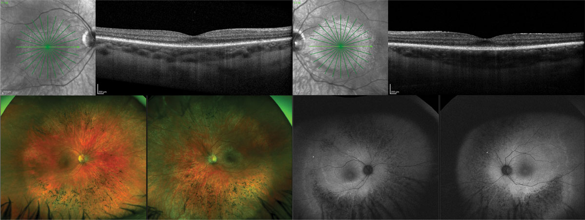
| Despite experiencing structural decline over the course of 10 years, several participants in this study with both autosomal recessive and autosomal dominant rod-cone dystrophy also maintained stable visual function. This highlights the need to further examination of distinct patterns of decline within and between genetic subgroups. Photo: Jessica Haynes, OD. Click image to enlarge. |
“This highlights the need for further examination of distinct patterns of decline within and between genetic subgroups, which is crucial for modeling long-term progression,” the authors of the study wrote in their paper.
A total of 23 participants attended follow-up (mean age 63 years, 48% female), with 20 classified as having rod-cone dystrophy and three re-classified as having cone-rod dystrophy based on genetic diagnosis. In advanced disease, both dystrophies show extensive photoreceptor loss throughout the retina, making it difficult to distinguish between the initial diagnosis of a rod-dominant or cone-dominant phenotype.
At 10-year follow-up, only 60% of RCD participants showed progression of ≥15 letters in either or both eyes, and 40% did not meet the criteria in either eye. Between the eye with poorer versus better VA at baseline, high symmetry in disease progression was observed for Goldmann visual field (VF), and moderate interocular symmetry in disease progression was observed for VA and ellipsoid zone (EZ) width. Baseline values influenced progression for VA and percentage change in Goldmann VF area, whereas total percentage change in EZ width did not differ across baseline values.
“Based on these findings, it could be speculated that a greater level of symmetry in progression, when measuring across a wide spectrum of disease stages, may be achieved using global function assessments, such as VF area,” the researchers noted. “To better understand the impact of inter-eye asymmetry, larger longitudinal studies are needed to explore factors influencing differences in progression rates between eyes.”
| Click here for journal source. |
Britten-Jones AC, Luu CD, Jolly JK, et al. Longitudinal assessment of structural and functional changes in rod-cone dystrophy: a 10-year follow-up study. Ophthalmol Sci. November 5, 2024. [Epub ahead of print]. |

