 |
History
A 10-year-old white female, accompanied by her parents, presented to the clinic with foreign body sensation and a “growth” in her right eye, superior temporally. The patient had just recently undergone strabismus surgery to help correct a small constant right exotropia. The growth started a couple of weeks prior and had been rapidly growing over the past week, according to her father. The patient had no remarkable systemic pathology, no known drug allergies, no other contributory history and took no medications.
Diagnostic Data
Her best-corrected entering visual acuity was 20/20 OD and 20/20 OS, through a mild, myopic astigmatic correction. External examination revealed a non-tender, red, pedunculated, soft, nodular papule on the right superior temporal palpebral conjunctiva. All other external findings were normal with no evidence of afferent pupil defect. The balance of anterior segment structures and anterior chamber was normal. Applanation pressures measured 15mm Hg OD and 13mm Hg OS. Dilated fundus examination was unremarkable in both eyes.
Your Diagnosis
Does this case require any additional tests? What does this patient’s history and clinical findings tell you about her likely diagnosis?
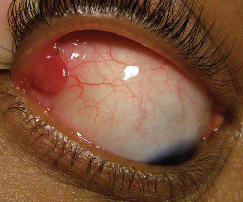 |
| The “growth” in this young patient’s right eye developed following strabismus surgery. Could the two be related? What’s your diagnosis? |
Discussion
Additional studies included manipulating the lesion, checking for hidden vascularity, firmness, movability, size and depth. The lesion was photo-documented and drawn with horizontal and vertical measurements.
The diagnosis in this case is a palpebral conjunctival pyogenic granuloma (PG). In the largest-published series of studies for vascular conjunctival tumors, PG was the most common, accounting for 22% of all reported cases.1 This lesion’s name is actually a double misnomer. First, the term “pyogenic” is defined as involving or relating to the production of pus; and second, “granuloma” is defined as a mass of granulation tissue.2-4 In fact, this condition is not caused by an underlying bacterial infection and usually lacks giant cells.1-4 Pyogenic granulomas have also been referred to as lobular capillary hemangiomas.2-4
Pyogenic granulomas occur secondary to a proliferative fibrovascular response. They present unilaterally in most instances with a slight male predilection.1-3 Peak incidence occurs in the third decade of life.1-3 Growth rate is rapid, (over days to weeks) and occurs in response to a provoking tissue insult, and in some cases during pregnancy.1-3 These events can range from trauma secondary to strabismus surgery (appearing at or near the muscle insertion) to procedures such as penetrating keratoplasty, scleral buckle placement and transconjunctival blepharoplasty.1-4 Other provoking traumas include chemical burns, the process of phthisis bulbi, inflammatory conditions such as chalazion and exposure or injury from ocular foreign bodies (such as an improperly installed punctal plug).1-3 Pyogenic granulomas may also be drug induced and are commonly prone to bleeding.1-3
Specifically, the lesion is composed of a profusion of immature capillary channels within a mixoid stroma containing: lymphocytes, leukocytes, fibroblasts and plasma cells. The surface is frequently bared of epithelium.1-4 A pyogenic granuloma usually presents as a fleshy red-pink mass with a florid blood supply from the adjacent conjunctiva.3-4 The lesion is often pedunculated, polypoidal and broadly based with an underlying stalk of feeder blood vessels and connective tissue.1-3 The condition is hypothesized to result from an imbalance in angiogenic regulation and is rarely idiopathic.1-5 Researchers have demonstrated that pyogenic granulomas are pathway-driven tumors, illustrating they may also arise from port-wine-stain-forming dermatologic and systemic conditions.6 Differential diagnosis for pyogenic granulomas includes Kaposi's sarcoma, Basal cell carcinoma, Squamous cell carcinoma, congenital haemangioma, glomous tumor.1-4 Pigmentation and color change are also marked signs to help distinguish benign vascular neoplastic lesions from other types of growths or tumors.1-3, 5
Pyogenic granulomas are benign lesions that usually respond to a short course of topical corticosteroid eye drops/ointments or local steroid injection.1-3,7 In most cases, surgical excision, cryosurgery, curettage, cauterization or laser removal are successful in proper removal of the lesion.1-4,7 The lesion has a tendency to recur locally upon surgical removal.1-3,7 Upon recurring episodes or continued growth, low-dose brachytherapy with a radioactive plaque has been proven effective.1-3,7 Follow up times of three to six months should be set to ensure stability with treatment.1-3,7
Dr. Gurwood thanks Christopher T. Luft, OD, for contributing this case.
|
1. Shields J, Mashayekhi A, Kligman B, et al. Vascular Tumors of the Conjunctiva. Ophthalmology. 2011; 118(1): 1747-1753. 2. Warner M, Bhat P, Jakobiec F. Subepithelial Neoplasms of the Conjunctiva. In: Warner M. Cornea, 3rd Ed. St. Louis: Mosby Elsevier; 2011: 485-507. 3. Shields J. Tumors of the Conjunctiva. In: Shields J. Shields C. Eyelid, Conjunctival, and Orbital Tumors: An Atlas and Textbook. 1st Ed. Philadelphia: Lipincott Williams & Wilkins; 2011: 350. 4. Wolters K. Conjunctival tumors. In: Gerstenblith A, Rabinowitz A, Barahimi B et al. Wills Eye Manual. 6th Ed. Philadelphia: Lippincott Williams & Wilkins; 2012: 130. 5. Lim Y, Douglas S, Ko C, et al. Somatic Activating RAS Mutations Cause Vascular Tumors Including Pyogenic Granuloma. J Invest Dermatol. 2015;135(6):1698-700. 6. Groesser L, Peterhof E, Evert M, et al. BRAF and RAS Mutations in Sporadic and Secondary Pyogenic Granuloma. J of Inv Dermatol. 2016;136(2):481-486. 7. Giblin A, Clover A, Athanassopoulos A, et al. Pyogenic granuloma: The Quest for Optimum Treatment. J Plast Reconstr Aesthet Surg. 2007;60(9):1030. |
