 |
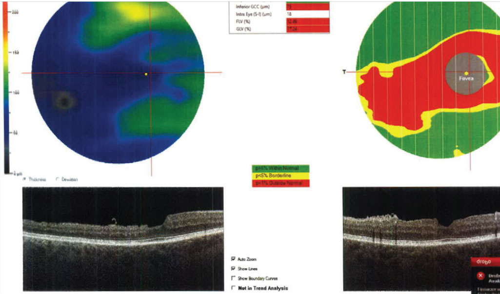
| Lower baseline superficial—but not deep—parafoveal vessel density was correlated with ganglion cell complex thinning in this study. Photo: Julia Reimold, OD. Click image to enlarge. |
One part of the eye favorably affected by glaucoma is the ganglion cell complex (GCC), and its thinning has been shown to correlate with change in visual field. Because of this, researchers decided to investigate a possible relationship between GCC thinning and baseline deep and superficial macular vessel density, hypothesizing that the latter would better detect glaucomatous change due to closer proximity to retinal ganglion cells and their axons found in the inner retina. Lower baseline superficial parafoveal vessel density (pfVD), but not deep pfVD, was associated with faster parafoveal GCC (pfGCC) thinning in primary open-angle glaucoma (POAG). They also found that superficial macular VD may help predict central macula thinning in patients with glaucoma.
The study compared 63 eyes of POAG patients with 34 eyes of glaucoma suspects, all of whom completed a minimum of four visits and two years of follow-up (n=69 patients). Both OCT and OCT angiography 3mm2 x 3mm2 macular scans were obtained at each visit and used to calculate superficial and deep pfVD and pfGCC thickness.
The baseline mean visual field mean deviation was -2.9 dB. An analysis of the univariable model landed researchers on evidence to support their hypothesis. “A lower baseline superficial pfVD and higher mean intraocular pressure (IOP) during follow-up were significantly associated with a faster pfGCC thinning rate, while deep pfVD was not,” the team noted. In addition, they observed in the multivariable model that faster pfGCC thinning correlated to higher mean IOP during follow-up and lower baseline superficial pfVD.
“Furthermore, eyes with a baseline superficial pfVD ≤46% (the lowest tertile) had a significantly faster pfGCC loss compared with eyes with a baseline superficial pfVD greater than 46%,” the researchers noted.
The study concluded that its findings demonstrate that “superficial capillary plexus, as compared with deep capillary plexus, is preferentially affected in glaucoma, and baseline superficial macular VD may be helpful to predict central macular GCC thinning.” The researchers also noted that “as central macula loss is directly related to central visual field defects and can negatively affect patients’ quality of life, information about superficial macular VD can help clinicians to identify high-risk patients who need more intensive observation and treatment.”
Wu JH, Moghimi S, Nishida T, et al. Correlation of ganglion cell complex thinning with baseline deep and superficial macular vessel density in glaucoma. Br J Ophthalmol. January 31, 2022. [Epub ahead of print]. |
