 |
|
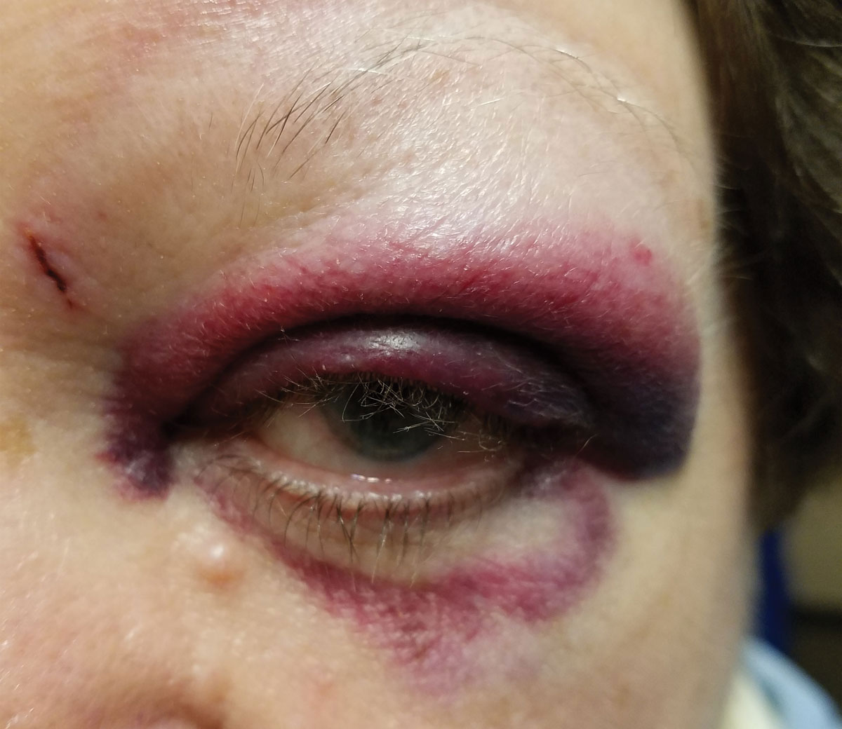
The most common ocular trauma findings reported were eyelid ecchymosis and edema (32% each), subconjunctival hemorrhage (42%), corneal laceration (32%), anterior chamber hyphema (56%), iris prolapse (18%), traumatic cataract (13%), vitreous hemorrhage (19%), optic neuropathy (3%) , macula commotio (5%) and retinal detachment (4%). Photo: Paige Thompson, OD. Click image to enlarge. |
One of the most common causes of permanent vision loss in adults is ocular trauma, with reports indicating that over one-quarter of cases (27%) end up with final visual outcomes that meet the legal definition of blindness. In order to accurately examine ocular trauma, physicians can use different ancillary imaging modalities, such as a CT scan, B-scan ultrasonography and/or ultrasound biomicroscopy (UBM), as direct examination is often hindered in trauma cases. To better understand ocular trauma in an adult population, and in particular the prognostic role played by imaging findings, researchers from the Cleveland Clinic attempted to find how examination and imaging findings correlate with treatment course and visual acuity outcomes. Their results were published recently in Journal of Ophthalmology.
In a retrospective chart review, the researchers collected the electronic medical records of 134 patients (71% male, 29% female) at a single institution from 2013 to 2020. Each patient had to have a history of either undergoing a CT scan (66%), B-scan ultrasonography (97%) and/or UBM (10%). A total of 136 unique ocular traumas were reported in this study.
The researchers reported on the presenting features and outcomes of their adult population. The median presenting visual acuity amongst the population was 2.7 logMAR with 62% of patients appearing with open globe injuries. Different ocular findings were found to be more prevalent depending on the imaging modality used on the patient. Globe deformity was the most prevalent amongst CT scan patients (30%), choroidal detachment was the most prevalent amongst B-scan ultrasonography patients (20%) and intraocular foreign body, ciliochoroidal effusions or angle recession were the most prevalent amongst UBM patients (21% each).
Patients who underwent B-scan ultrasound presented with the most significant data. Those who were positive for retinal detachment appeared to have worse vision on initial B-scan compared to patients who were negative for this finding at six months and at final post-injury evaluation. Furthermore, patients with choroidal detachment appeared to have worse visual acuity (VA) on initial B-scan compared to patients without this finding at six months and at final post-injury evaluation.
“In the ophthalmic population, B-scan imaging is routinely utilized to evaluate the globe when the view to the fundus is obstructed by anterior segment pathology or other media opacities,” commented the researchers in their study. “These same factors likely influence long-term visual acuity as found in this population; unsurprisingly, positive findings on B-scan were associated with worse visual acuity at follow-up.”
Zone of ocular injury was defined according to the location of the most posterior full-thickness aspect of the globe opening for open globe injuries or most posterior identified pathology for closed-globe injuries, the researchers wrote in their paper. Zone I was limited to the cornea or corneoscleral limbus, Zone II involved the anterior 5mm of the sclera and Zone III denoted at a location more than 5mm posterior to the corneoscleral limbus.
The median logMAR visual acuity at the six-month post-injury time point was 0.7 and at the final post-injury VA was 0.5 logMAR. There was no significant difference in VA at six-month or final postoperative evaluation between patients with different zones of injury, regardless of open or closed globe status, the paper describes. “However, in the open globe category, there was a general trend towards worse vision with more posterior zones of injury (zones 2 and 3),” the authors wrote. The presence of positive exam findings in the conjunctiva/sclera and optic nerve were associated with worse VA at six months and final post-injury status, while positive findings in the vitreous and the periphery were associated with better VA at the final post-injury evaluation.
Since this was a retrospective study limited to data at a single institution, the researchers recognize that the impact of their results is limited. One potential limitation was the use of past documentation that could have been gathered by clinical residents and fellows rather than an experienced practicing physician. Additionally, six-month and final post-injury follow-ups were observed in the study, but 25% of the patient population did not attend their six-month follow-up visit. While this may have skewed the results, the researchers noted that 98% of the patient population did attend their final post-injury follow-up visit.
“Further investigation with a prospective study where all patients receive the same ancillary tests would be needed to determine if specific imaging findings are truly predictive of final outcomes and an eventual need for secondary surgery,” concluded the researchers in their study.
Clevenger LM, Cao JL, Steinkerchner MS, et al. Demographics, presenting features, and outcomes of adult patients with ocular trauma. Journal of Ophthalmology. May 29, 2024. [Epub ahead of print]. |
