Spectral-domain optical coherence tomography (SD-OCT) is widely adopted for precise RNFL thickness measurements and other optic nerve head parameters and has become the standard of care for imaging in glaucoma patients. A recent study has provided evidence for racial differences in the diagnostic accuracy of RNFL thickness in glaucoma detection, being significantly lower in African descent compared with European descent eyes. The extent of the differences varies with the specific OCT device used for testing and was more robust with the Spectralis (Heidelberg Engineering) than the Cirrus (Carl Zeiss Meditec).
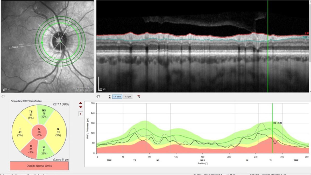 |
| The devices used in this study have inherent limitations in their ability to image the sclera and lamina cribrosa, which may have provided clues for such racial differences. It is also important to note that, while the mean deviation was similar between the groups in the present study, the exact onset of glaucoma is not known and may have confounded the results. Photo: James Fanelli, OD. Click image to enlarge. |
The researchers included 379 healthy eyes (125 African descent and 254 European descent) of 209 patients and 442 glaucomatous eyes (226 African descent and 216 European descent) of 340 patients from the Diagnostic Innovations in Glaucoma Study and the African Descent and Glaucoma Evaluation Study. Glaucoma was defined as the presence of glaucomatous visual field damage confirmed on three consecutive and reliable standard automated perimetry 24-2 tests. Abnormality was defined as pattern standard deviation of ≤ 5% and/or glaucoma hemifield test outside normal limits. Healthy eyes were defined as intraocular pressure (IOP) < 22mm Hg with at least two reliable, consecutive normal visual fields.
Diagnostic accuracy for Spectralis-RNFL thickness was significantly lower in eyes of African descent compared with those of European descent (area under the curve: 0.85 and 0.91). Results for Cirrus-RNFL thickness were similar but did not reach statistical significance (0.86 and 0.90 in African and European descent, respectively), which may be attributed to the smaller sample size in the Cirrus group. Adjustments for age, central corneal thickness, axial length, disc area, visual field mean deviation and IOP yielded similar results.
“The consistency of the obtained results after adjustment for many potential confounders suggests that the results are robust within the range of the parameters evaluated,” the authors wrote in their paper, which was published in American Journal of Ophthalmology.
Healthy eyes of African descent had a significantly thicker Cirrus RNFL thickness compared with those of European descent (mean values: 96.75µm and 93.29µm in African descent and European descent respectively). Furthermore, glaucomatous eyes of African descent had significantly thicker Spectralis (mean values: 80.2µm and 74µm in African descent and European descent respectively) and Cirrus-RNFL thickness (76.93µm and 72.84µm in African and European descent respectively.
The researchers proposed many reasons for the difference such as the tendency of individuals of African descent to have larger optic discs, and measurements obtained closer to the optic disc margin tend to be thicker. Another possible explanation was that the lower diagnostic accuracy of RNFL thickness in African descent eyes is a form of poor structural-functional correlation. In other words, individuals of African descent may experience visual function impairment earlier than RNFL thinning can be detected.
“The consistently lower diagnostic performance of RNFLT in detecting glaucoma in populations of African descent may indicate significant racial differences in the IOP-driven remodeling of the optic nerve head connective tissue, which includes the peripapillary sclera and the lamina cribrosa,” they wrote.
“Further longitudinal studies with enhanced imaging of the deeper optic nerve head layers may provide clues about the reasons underlying these differences, which could improve the utility of RNFL thickness in clinical practice and introduce new biomarkers for glaucoma detection,” the study concluded.
KhalafAllah MT, Zangwill LM, Proudfoot J, et al. Racial differences in diagnostic accuracy of retinal nerve fiber layer thickness in primary open-angle glaucoma. Am J Ophthalmol. October 28, 2023. [Epub ahead of print]. |