 |
| Techniques to measure arteriole (red) and venule (blue) diameter may one day become part of a protocol for cognitive assessment. Photo: Amy Jane Mcgowan/PLoS One. Click image to enlarge. |
Evidence suggests certain mid-life factors may be predictive of future neurodegenerative disease. Researchers recently investigated retinal vessel diameters and their association with brain aging (a sensory and cognitive composite measure) and macular ganglion cell inner plexiform layer (mGCIPL) thickness (a neurodegeneration biomarker) and reported that vessel measurements may offer clues to neurodegeneration and risk assessment.
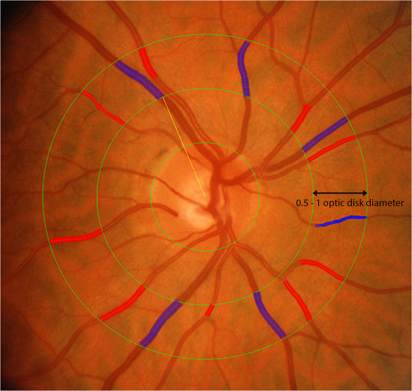
The researchers quantified vessel diameter using measurements called central retinal arteriolar and venular equivalents (CRAE and CRVE) from fundus images of Beaver Dam Offspring study participants (n=2,381). Mean CRAE and CRVE were 148.8µm and 221.7µm, respectively. The team found an association between CRAE and brain aging among participants without ocular pathology, where wider CRAE was associated with a 33% decreased risk of brain aging over five years. Additionally, CRAE and CRVE were both independently associated with mGCIPL thickness. The researchers reported that mGCIPL thickness increased by about 1.7µm and 2.0µm per SD increase in CRAE and CRVE, respectively.
“Among those without known eye disease, having wider CRAE was associated with lower brain-aging risk,” the researchers concluded in their paper. “CRAE may serve as a screening tool, identifying those at higher risk for cognitive and sensorineural decline among those without clinically identified ocular pathology.”
Paulsen AJ, Pinto AA, Merten N, et al. Association of central retinal arteriolar and venular equivalents with brain-aging and macular ganglion cell-inner plexiform layer thickness. Ophthalmic Epidemiol. March 28, 2022. [Epub ahead of print]. |
