Researchers continue to uncover ocular evidence of neurodegenerative diseases, potentially laying the groundwork for future diagnostic methods. In the largest cohort of Parkinson’s disease (PD) patients studied to date, new research centered on identifying retinal vascular characteristics using ultra-widefield (UWF) scanning laser ophthalmoscopy. UFW imaging was used in both PD patients and controls with normal cognition and no PD. Excluded were patients with diabetes, uncontrolled hypertension, glaucoma, dementia, other movement disorders or known retinal or optic nerve pathology.
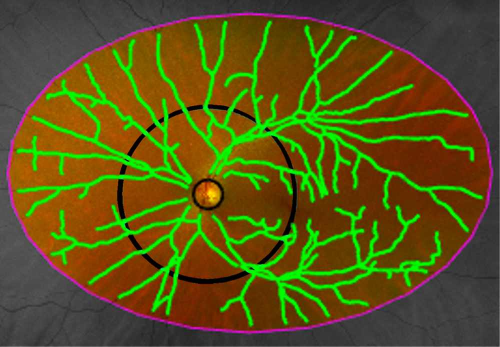 |
| This study used a conventional Optos UWF imaging device to capture images, which were then analyzed by custom software to segment retinal vessels. The researchers manually selected the largest and longest artery and vein pair within each quadrant (superotemporal, inferotemporal, inferonasal, superonasal); vessel widths and tortuosity were extracted from these annotated vessel paths. Photo: Ma JP, et al. Transl Vis Sci Technol. 2024;13(1):15. Click image to enlarge. |
The custom software used by the researchers described retinal vessel width and tortuosity and included other capabilities to further characterize vascular morphology. The PD cohort included 53 eyes of 38 individuals; the controls included 51 eyes of 33 individuals. Eyes of patients with PD displayed more tortuous retinal arteries in the superotemporal quadrant, as well as retinal vascular spread, when compared with controls. However, analysis suggested increased vascular network complexity in those with PD.
Expanding further on their data, the study authors continue that no findings indicated differences in measurement of vessel width gradients, echoing previous research which measured vessel caliber with OCT in PD patients vs. age- and sex-matched controls.
They also point to their finding of greater arterial tortuosity in the superotemporal quadrant of PD eyes. There was another, similar trend toward greater tortuosity in the superonasal arteries, pointing to a potential for the superior retina to be an area of research in future studies. Vascular tortuosity measurement as a cellular stress marker in the retina is an emerging area of research, as is understanding retinal vascular tortuosity’s mechanism and pathophysiology as it relates to PD. It is still unclear if vascular tortuosity in the retina is a primary or secondary phenomenon in PD.
The researchers found data supporting both decreased spread of retinal vessels in PD and increased complexity of a retinal vascular network in PD eyes. This variant of normal morphology may indicate a marker of vascular stress secondary to neurodegeneration. The authors speculate early microvascular change may be caused by early exhaustion of capillary beds from PD pathophysiology. Microvascular change may then be followed by macrovascular retraction.
Overall, the results of this study are clinically relevant, the researchers wrote, because “retinal vasculature assessment using these novel methods may be useful in understanding ocular manifestations of PD and the development of retinal biomarkers.”
Ma JP, Robbins CB, Pead E, et al. Ultra-widefield imaging of the retinal microvasculature in Parkinson disease versus controls with normal cognition using alpha-shapes analysis. Transl Vis Sci Technol. 2024;13(1):15. |
