 Click image to enlarge. Click image to enlarge. |
Corneal crosslinking (CXL) is a common procedure used to treat progressive keratoconus that can potentially halt corneal thinning and steepening to preserve vision. Though this treatment has a very high success rate overall, there are multiple factors that play into a patient’s likelihood of achieving an ideal visual outcome post-surgery. According to new research presented by Philip W. Dockery, MD-MPH, during the 2022 ASCRS annual meeting this past weekend, a patient’s race may also be predictive of treatment success. In fact, Dr. Dockery’s paper—a retrospective analysis of 508 eyes that underwent CXL for keratoconus—concluded that treatment failure (repeat CXL or corneal transplant) was 4.5 times more common among Black patients than white patients after controlling for demographics and medical history.
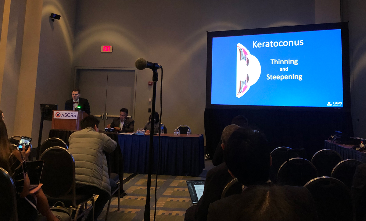 |
| Dr. Philip Dockery presenting his team's paper on clinical outcomes of CXL during this year's ASCRS meeting. Click image to enlarge. |
Dr. Dockery and his team performed CXL on the cohort (257 white patients and 251 Black patients) and then analyzed the data to determine patterns in clinical presentation, complications and outcomes. The team evaluated factors such as demographics, comorbidities including allergies and habits such as eye rubbing. They also sought to identify potential predictors of treatment failure.
The analysis supported other research showing that treating keratoconus patients with CXL overwhelmingly results in positive clinical outcomes. “What we found is that crosslinking is fantastic; it has incredible rates of halting progression,” said Dr. Dockery during his presentation. “After one year, 97% of patients showed no signs of progression, and after two years, 91% of patients showed no signs of progression.” He noted that only one patient in the entire study required a corneal transplant.
The main factor that the team concluded had contributed to the higher rate of treatment failure in Black patients was the difference in disease presentation between the two populations. Compared with white patients, on average, Black keratoconus patients in the study who underwent CXL were found to have more progressive disease, a thinner central corneal thickness and a steeper K1, K2 and Kmax (Table 1). The only significant demographic differences between Black and white patients were that more white keratoconus patients were male (74% vs. 39%) and fewer reported eye rubbing (73% vs. 83%).
“We know that advanced keratoconus is more likely to lead to progression,” Dr. Dockery told attendees of his presentation. “Our study showed that more advanced disease presentation and more negative treatment outcomes were both observed among Black patients in our study. So, why could this be? It could be because there’s a disparity in access to care leading to delayed presentation in these individuals,” he explained.
Dr. Dockery wrapped up his talk by emphasizing the need for more efforts promoting earlier disease detection and intervention. “These findings highlight the importance of early crosslinking as well as developing collaborative networks with the patient’s primary eye physician to be able to catch the disease early and optimize outcomes,” he concluded.
Table 1. Corneal Measurements in Black vs. White Keratoconus Patients
Average Pre-op Measurements | Black Patients (n=251) | White Patients (n=257) |
CCT | 462μm | 488μm |
K1 | 50.5D | 46.5D |
K2 | 55.1D | 50.4D |
Kmax | 63.8D | 57.8D |
