 |
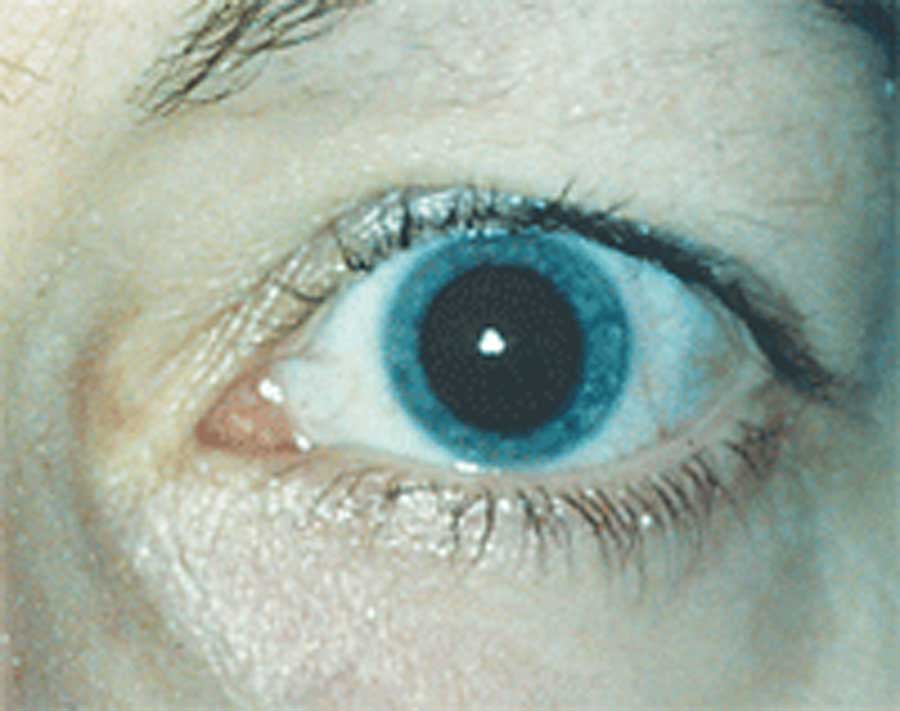
Manual eyelid lifting and pupil dilation may allow you to obtain a clearer, more visible scan of the retina. Photo: Ellen M. Petrilla, OD. Click image to enlarge. |
Maximizing visualization of the peripheral retina is important for optimal characterization of diabetic retinopathy (DR) severity. Even with ultra-widefield (UWF) retinal imaging, there can be significant variability in the extent of visible retinal area between images. Some of this variability is due to artifacts resulting from the patient’s eyelashes and eyelids. Researchers recently determined that pupillary dilation and eyelid lifting can substantially increase the visual retinal area and detect predominantly peripheral lesions and hemorrhages and/or microaneurysms associated with DR risk.
The study used fully automated visible retinal area and hemorrhage and/or microaneurysm count detection algorithms on 5,919 eyes from 3,014 participants (43.2% female, 81.3% Caucasian, mean diabetes duration: 15.9 years) from two cohorts, one with non-mydriatic UWF imaging from a DR teleophthalmology program and one with mydriatic imaging from an academic retina practice. Predominantly peripheral lesions and hemorrhages/microaneurysms were defined as present when at least one field had greater hemorrhage and/or microaneurysm number in the peripheral retina than within the corresponding ETDRS field.
The researchers found that dilation increased visible retinal area by 25% and simple eyelid lifting increased the area by an additional 10%. Manual eyelid lifting improved visible retinal area by 11% in the undilated eye. Increased visible retinal area was associated with detecting increased numbers of hemorrhages and/or microaneurysms and a greater likelihood of identification of predominantly peripheral lesions. Furthermore, predominantly peripheral lesions were more common in all DR levels with mydriatic imaging compared with non-mydriatic imaging. The discrepancies suggest that, without mydriasis, these lesions and other DR lesions may be missed.
“Given the importance of hemorrhages, aneurysms and predominantly peripheral lesions for determining risk of DR progression, these findings emphasize the importance of maximizing visible retinal area in addition to image quality for optimal risk assessment in clinical trials and teleophthalmology programs,” the study authors concluded.
Jacoba CMP, Ashraf M, Cavallerano JD, et al. Association of maximizing visible retinal area by manual eyelid lifting with grading of diabetic retinopathy severity and detection of predominantly peripheral lesions when using ultra-widefield imaging. JAMA Ophthalmol. February 24, 2022. [Epub ahead of print]. |
