Although photorefractive keratectomy has been overshadowed by LASIK, PRK still is performed routinely today. In the 18 years since its FDA approval, PRK has been improved by use of larger treatment zones, smoother ablations and immunomodulatory drugs. Such upgrades have yielded increased accuracy. In fact, PRK has stood the test of time so well that many surgeons now refer to it as “advanced surface ablation.”
Ironically, the benefit of PRK can also sometimes be its biggest deterrent. If, for example, patients have epithelial or anterior stromal irregularities––such as epithelial basement membrane dystrophy or anterior stromal scars from ulcers––the diseased tissue is removed during the procedure. (LASIK, by contrast, would preserve these defects.) Unfortunately, however, the epithelial regrowth phase may last several days or weeks, resulting in a prolonged post-op period of reduced visual acuity.
Usually, surgeons choose to loosen the epithelium with alcohol. This is accomplished by first creating a well in the corneal tissue, which is then filled with an alcohol solution. The loose epithelium is gently removed with a sterile surgical spear, permitting direct laser access to the stroma, which is then cleaned and gently polished with a blade.
During the ablation, the patient fixates on a central light. A flying spot pattern is used to prevent tissue overheating and to increase accuracy. Typically, the surgeon will apply a sponge soaked with mitomycin C to the cornea for a specified duration to decrease the risk of postoperative haze. The eye is then rinsed with buffered saline to remove any residual debris. Finally, the cornea is covered with a bandage lens.
Multimedia
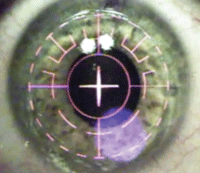
While the patient fixates on a central light, a flying spot pattern is used to minimize tissue heating and to increase accuracy. Click
here to see a video of this procedure being performed.
Postoperatively, patients are usually seen the next day to ensure that the bandage contact lens remained in place. A large epithelial defect will be visible at this follow-up.
The patient should then be scheduled for a second follow-up five to seven days after surgery. Typically, re-epithelialization will be complete within a week. At this time, the bandage contact lens may be removed. If any loose or incomplete epithelium remains, the lens may be left in place for several additional days. During the subsequent month, patients should taper postoperative steroid drops while enjoying progressively better vision.
When counseling patients, optometrists should:
- Emphasize the extended visual recovery period compared to LASIK, and reassure the patient while he or she is healing. No matter how adequately prepared you believe patients are for slow visual recovery, they can become extraordinarily concerned when they do not see well after a short time.
- Prepare patients for photophobia, discomfort and pain. Discomfort typically persists for just two days after surgery. Again, forewarned is forearmed. With the routine use of topical NSAIDs and bandage lenses, very few will report significant pain. Having no way to predict which patients will or will not experience significant pain, we always prescribe a three-day course of oral narcotics.
