 |
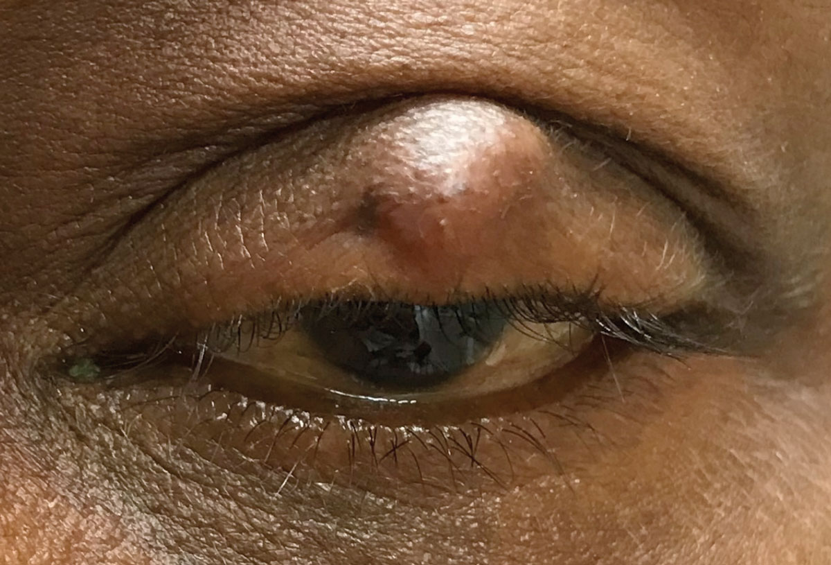
Bacterium C. acnes may play a role in chalazion development. Photo: Joseph W. Sowka, OD. Click image to enlarge. |
It’s been reported that patients with meibomitis-related keratoconjunctivitis (MRKC) have a history of chalazion and that MRKC’s bacteria is considered to be Cutibacterium acnes (C. acnes). In a recent study, researchers evaluated the clinical characteristics of chalazion cases and performed immunohistochemical examinations of chalazion tissue samples following surgery to investigate any possible etiological link between chalazion and C. acnes.
The age and sex of 206 Japanese patients of Asian ethnicity (124 females, 82 males) with chalazion were examined. A total of 28 curettage samples obtained at the time of surgery were fixed in 10% buffered formalin, paraffin-embedded, thinly sliced and subjected to hematoxylin and eosin staining. Of those, 20 were histologically confirmed as chalazion, while the rest were confirmed to be granulation tissues.
The serial sections further underwent immunohistochemical examination utilizing an automated system with a commercially available C. acnes-specific monoclonal antibody (PAB antibody) that reacts with membrane-bound bacterial lipoteichoic acid.
Among the 20 histologically-confirmed chalazion cases, seven were positive for PAB antibodies. PAB (+) cells were often seen with clusters of neutrophils in the granulomas consisting of epithelioid cells and lymphocytes, indicating immune granulomas accompanied by neutrophils.
In contrast, typical PAB (-) granulomas often contained lipid vacuoles in multinucleated giant cells surrounded by lymphocytes and histiocytes rather than epithelioid cells, thus indicating lipogranulomas. Three of the seven PAB (+) cases (all males) had multiple chalazia in more than one eyelid at initial presentation.
“The findings in this study are the first to reveal positive staining for C. acnes in the granulomas of chalazion, implying a possible role for C. acnes in granuloma formation,” the authors explained. “Moreover, the chalazion tissues with positive reactivity to PAB antibody showed epithelioid cell granulomas accompanied by neutrophil infiltration, suggesting an immune-type granulomatous reaction to C. Acnes bacterium.”
The findings also revealed that the prevalence of chalazion was significantly higher in females 40 years and younger, which suggests it’s more common in premenopausal women.
“In summary, our study shows the possible involvement of C. acnes and female sex hormones in the pathogenesis of chalazion,” the authors concluded. “Although chalazion has been well accepted as a foreign body reaction to degenerated meibum (“lipogranuloma”), C. acnes may be involved in granuloma formation in some cases, especially those with conspicuous epithelioid cells and neutrophil infiltration. Additional immunohistological examinations are needed to fully elucidate the pathogenesis of chalazion.”
Suzuki T, Katsuk N, Tsutsumi R, et al. Reconsidering the pathogenesis of chalazion. Ocul Surf. December 20, 2021. [Epub ahead of print]. |
