Early detection and accurate assessment of glaucomatous damage are crucial for effective management and preservation of visual function. Optical coherence tomography (OCT) and visual field testing are widely used in glaucoma evaluation, but they are limited in their ability to precisely identify and correlate structural and functional abnormalities, particularly in the macula. New research published in American Journal of Ophthalmology aimed to enhance the understanding and detection of glaucomatous damage in this region by investigating the association between papillofoveal or papillomacular bundle defects and abnormalities in 10-2 visual field sensitivity using a novel imaging processing algorithm.
The technology used was called RNFL optical texture analysis (ROTA) to provide detailed trajectorial information of axonal fiber bundles within and beyond the macula that the researchers note may go unnoticed in OCT RNFL thickness analysis and red-free RNFL photography. “Using widefield OCT scans including peripapillary and macular regions, it becomes possible to map individual visual field stimuli to their topographically corresponding locations on the ROTA map, enabling a more detailed assessment of structure-function relationship at the pointwise level,” the researchers explained in their AJO paper.
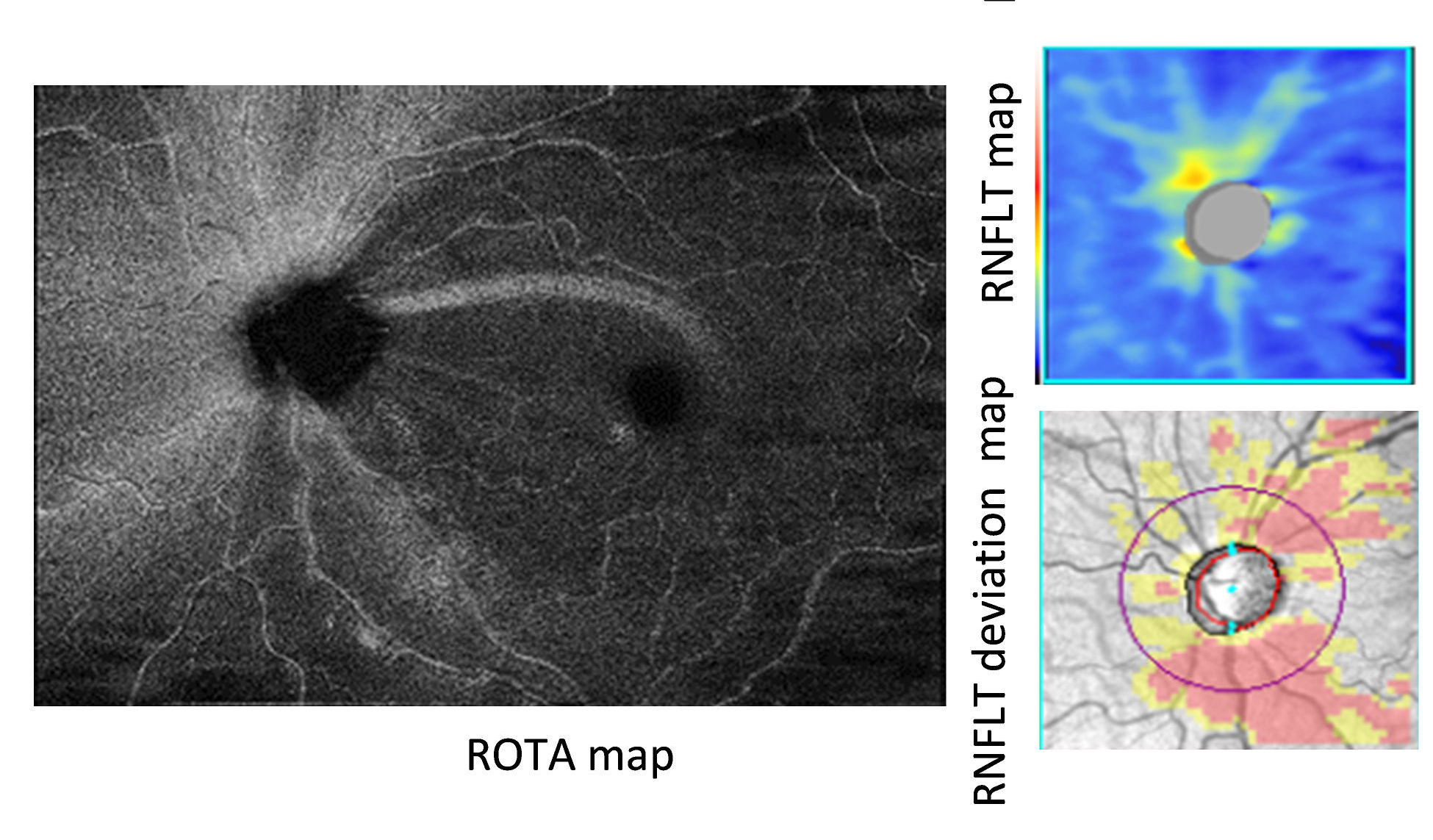 |
| Papillomacular and papillofoveal RNFL bundle defects were a common finding in eyes with glaucoma in this study with prevalence rates of 92.1% and 37.9%, respectively. Photo: Leung CK, et al. IOVS 2018;59:3497. Click image to enlarge. |
The study involved 841 eyes that were categorized as healthy (n=44), glaucoma suspect (n=317) and glaucoma (n=380). The prevalence of RNFL bundle defects and the involvement of the arcuate, papillomacular and papillofoveal bundles in each eye, as determined by ROTA, were assessed across the different groups, including healthy eyes, eyes with ocular hypertension, eyes with glaucomatous optic neuropathy and eyes with glaucoma of varying severities. All eyes also went underwent 24-2 and 10-2 visual field testing.
The data showed that papillomacular (92.1%) and papillofoveal (37.9%) RNFL bundle defects were both prevalent findings in eyes with glaucoma. Additionally, the researchers observed that a 10-2 VF location projected onto a papillomacular or a papillofoveal RNFL bundle defect had a significantly increased likelihood of reduced sensitivity (odds ratios: 18.61 and 20.17, respectively). “This provides evidence that the visualized papillomacular and papillofoveal bundle defects on ROTA are indeed precisely associated with central visual field defects,” they noted in their paper.
The imaging modality demonstrated predictive value in identifying 10-2 visual field abnormalities, with higher odds ratios compared to abnormalities in corresponding central locations of the 24-2 test. The study authors wrote that, “To predict the likelihood of visual field sensitivity abnormalities on the 10-2 test, involvement of the corresponding central 24-2 test location and the presence of papillomacular and papillofoveal RNFL bundle defects offered complementary insights, with the latter demonstrating a higher magnitude of the effect.”
Once ROTA is validated in real-world settings, the imaging modality may help provide valuable insights into the patterns of glaucomatous damage, especially within the macula, as well as play a role in the assessment of central visual field involvement.
Kamalipour A, Moghimi S, Khosravi P, et al. Retinal nerve fiber layer optical texture analysis and 10-2 visual field assessment in glaucoma. Am J Ophthalmol. May 24, 2024. [Epub ahead of print]. |
