In April 2016, optometry lost a giant when the author of the seminal work Primary Care of the Posterior Segment, Larry Alexander, OD, died. In addition to being an optometric physician, author and educator at the University of Alabama Birmingham School of Optometry, Dr. Alexander was a past president of the Optometric Retina Society (ORS). That group chose to honor his legacy by accepting case reports from optometric residents across the country relating to vitreoretinal disease.
The two cases shown here, selected by the ORS Awards Committee, were co-winners of the fifth annual Larry Alexander Resident Case Report Contest. The contest is sponsored by Zeiss, Heidelberg and Optos.
“When reviewing submissions, we look for manuscripts that are well written, include high-quality images and provide new and innovative information to the community,” says Julie Rodman, OD, professor and chief of the Fort Lauderdale (Broward) Eye Care Institute at Nova Southeastern University in Florida, and ORS treasurer. “This year, our winning manuscripts focused on timely topics and provided insight into the diagnosis and management of entities that are often challenging to diagnose,” she notes.
The full text and images of both case reports are available online at the links below.
Case 1: Choroidal Melanoma
This case study, presented by Amy Bade, OD, a low vision and ocular disease resident at the Northeastern State University College of Optometry, reviewed characteristic risk factors for choroidal melanoma and the use of multimodal imaging to assist in the detection of these key features.
The report detailed the management of a Native American woman diagnosed with a choroidal melanoma and discussed the findings and clinical utility of supplementary multimodal imaging. According to Dr. Bade, early and accurate detection of choroidal melanoma has greatly improved with the use of contemporary multimodal imaging technology, including enhanced-depth imaging OCT, fundus autofluorescence and widefield fundus photography.
 |
|
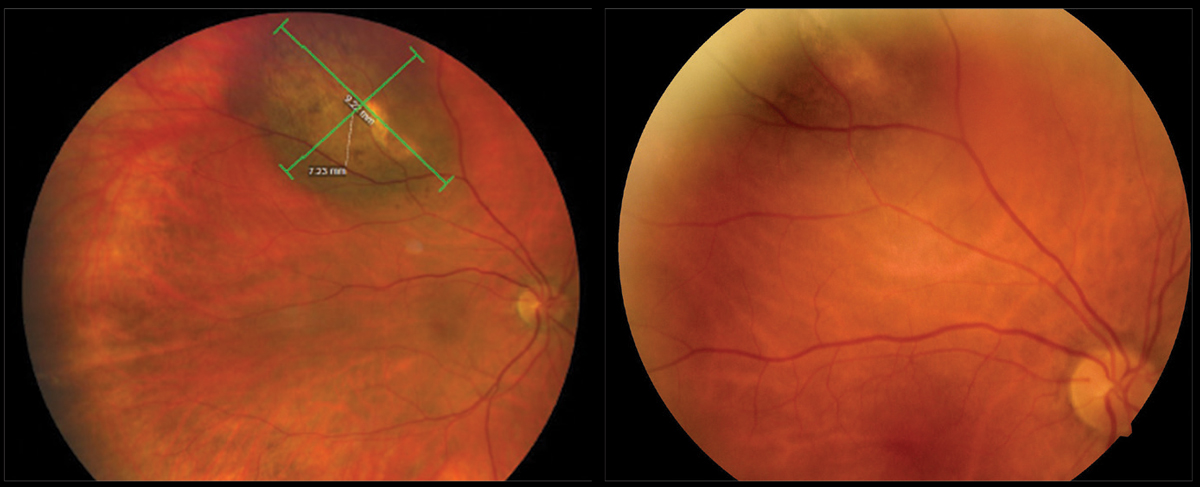
Comparing images from 2020 (left) and 2018 (right) of the pigmented choroidal tumor of the right eye demonstrates obvious expansion of the choroidal melanoma. Click image to enlarge. |
Contemporary management of choroidal melanoma most often includes plaque radiotherapy, and newer, innovative methods are promising. Although the condition is uncommon, Dr. Bade believes it is highly critical that eye care providers be familiar with and able to recognize high-risk features suggestive of choroidal melanoma by using multimodal imaging.
Case 2: Pediatric CHRRPE
 |
|
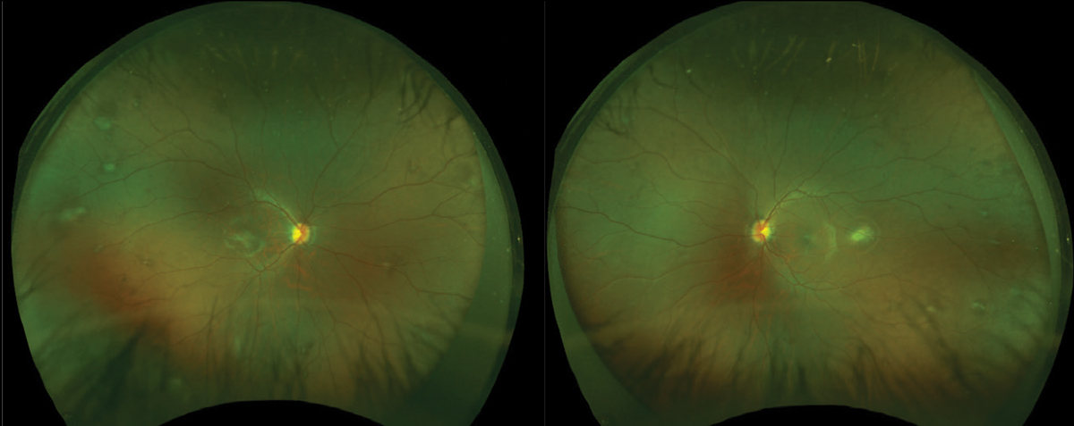
Fundus images of the right and left eyes reveal that CHRRPE lesions often appear elevated and pigmented (usually grey, white or brown, but may be orange, yellow or green). Click image to enlarge. |
In her case report, Dr. Topete discussed the clinical characteristics of CHRRPE, as well as other potential ocular manifestations of NF2 and the importance of early diagnosis. She noted that it is important for eye care practitioners to recognize that children with NF2 more commonly present with ocular, dermatological and/or neurological signs and require careful ophthalmic examination.
