Although age-related macular degeneration (AMD) is likely linked with dementias such as Alzheimer’s disease (AD) in some capacity, the results of this new study determine that, contrary to some associated reports, the status of the inner retina is not linked with cognition. The study researchers wanted to assess whether retinal layer thicknesses, including the retinal nerve fiber layer (RNFL), are associated with cognition in AMD. This was emphasized due to both AD and AMD affecting older adults and sharing risk factors. As well, AD and its precursor states demonstrate a thinner RNFL when compared with similarly aged controls.
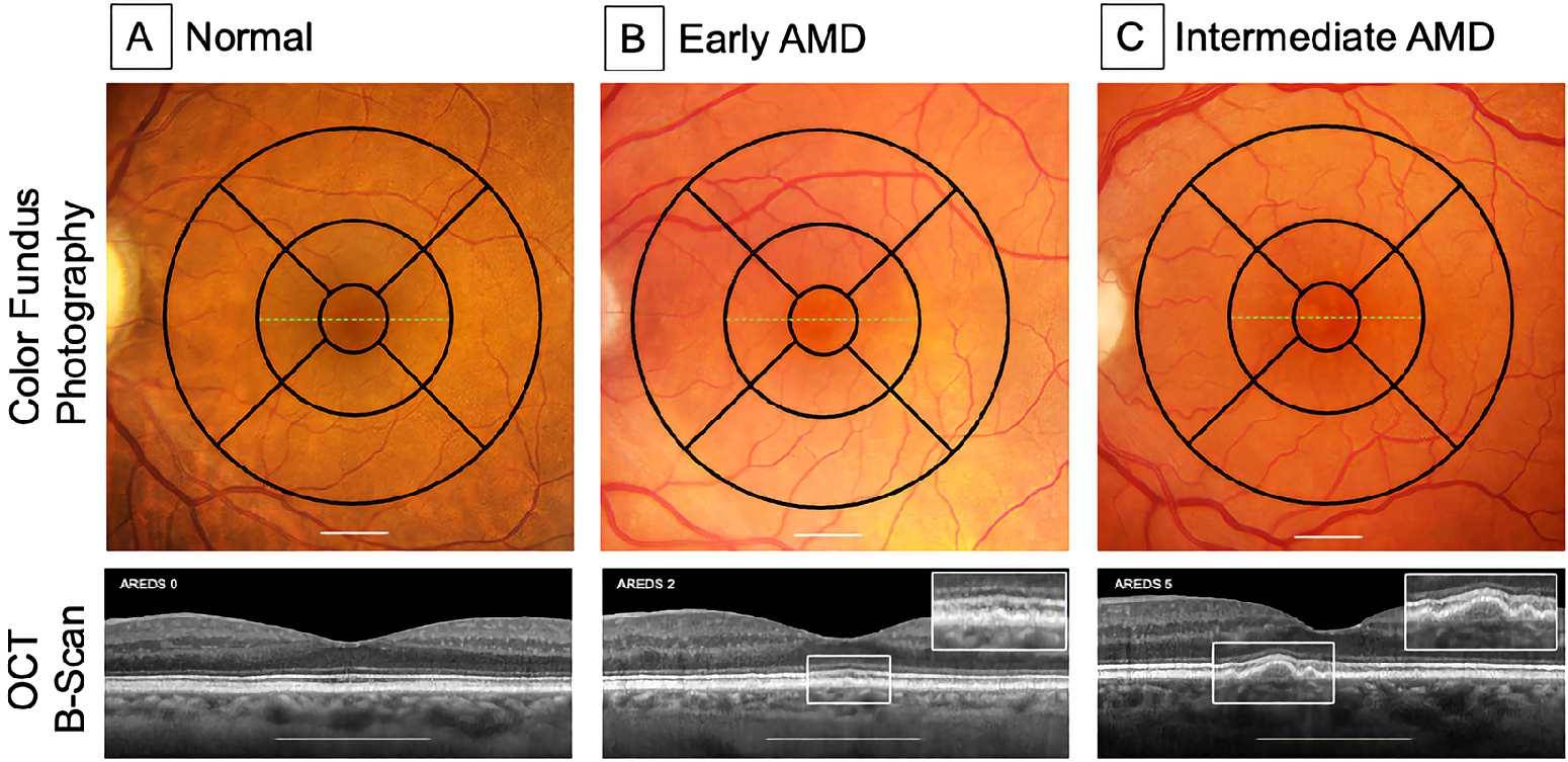
To elucidate the relationship, the authors included 63 total adults aged 70 or older with normal retinal aging, early AMD and intermediate AMD; 21 people were included in each group. OCT volumes underwent 11-line segmentation and adjustments by a trained operator. Evaluated thicknesses reflect the vertical organization of retinal neurons as well as two vascular watersheds: RNFL, ganglion cell layer-inner plexiform layer complex, inner retina, outer retina including retinal pigment epithelium to Bruch’s membrane and total retina.
 |
|
Some mechanisms that may impact the retina and the brain are microglia populations reflecting specific niches and phases of neurodegenerative disease, infiltration by circulating immune cells, long-term effects of prior infection and the impact of microbiota on inflammation and nutrient absorption. Photo: Owsley C, et al. Invest Ophthalmol Vis Sci. 2024;65(5):16. Click image to enlarge. |
The only notable result of correlations estimated for association between cognition and thickness was of the outer retina; thinning of this layer was significantly associated with lower cognition scores. None of the other layers’ thicknesses were associated with cognition.
Due to this observation, the authors note that “early and intermediate AMD are stages of a disease whose first-order and primary impact is in the outer retina and its supporting vasculature with functional changes in inner retinal layers occurring as circuitry eventually degrades.” The affected outer layer consists of photoreceptors, supporting glia, retinal pigment epithelium and Bruch’s membrane. The lack of RNFL association with cognition was in contrast with multiple AD-associated reports.
Consequently, this led the authors to ask how outer retinal thickness reductions could impact cognitive function, leaning into the idea that, “our findings may hint at a uniquely shared factor impacting brain and outer retina in persons with AMD whose outer retina is degenerating.”
As they explain, previous research has found thinning if the outer nuclear layer and ellipsoid zone in those with frontotemporal dementia compared with healthy controls, with no inner retinal layer thickness differences seen. As well, thinning of these layers mapped onto results of a Mini-Mental State Examination. The authors of this previous work suspected microtubule-associated defects, identifying outer nuclear layer thinning and ellipsoid zone defects in mice with a confirmed Rp1 mutation. RP1 and tau are both microtubule-associated proteins in photoreceptor cilia, making this a possible link.
With these authors soon coming out with two other investigations related to this one, they impart that, “together, these studies can serve as preliminary information to motivate hypotheses for a more comprehensive examination of the potential associations between AMD and AD, two of the most common and devastating neurodegenerations of aging.”
Owsley C, McGwin Jr. G, Swain TA, et al. Outer retinal thickness is associated with cognitive function in normal aging to intermediate age-related macular degeneration. Invest Ophthalmol Vis Sci. 2024;65(5):16. |
