Although not yet precise, a newer OCT-A metric gives clinicians and researchers a glimpse of future functional progression in the glaucoma patients. Researchers believe that rapid initial optic nerve head capillary density loss may be used to assess the risk of glaucoma visual field progression. Their recent study, published in JAMA Ophthalmology, found that rapid initial optic nerve head (ONH) capillary density loss from OCT-A was associated with a faster rate of visual field progression and a doubling of the risk of developing event progression. They believe that their method could potentially help identify patients who demonstrate a high risk for fast glaucoma progression, resulting in more severe functional impairment and potentially in blindness, which is paramount for optimizing management strategies.1
The retrospective study of a longitudinal cohort at a glaucoma referral center included 167 eyes (96 with primary open-angle glaucoma and 71 with glaucoma suspect) of 109 patients (mean age: 69.0; 51.4% female) who were monitored for a mean of 5.7 years. The rates of initial capillary density and average retinal nerve fiber layer loss were calculated from the first three optic nerve head OCT-A and OCT scans, respectively, during the initial follow-up (mean 2.0 years). Based on the median rate, eyes were categorized into fast- and slow-progressor groups.
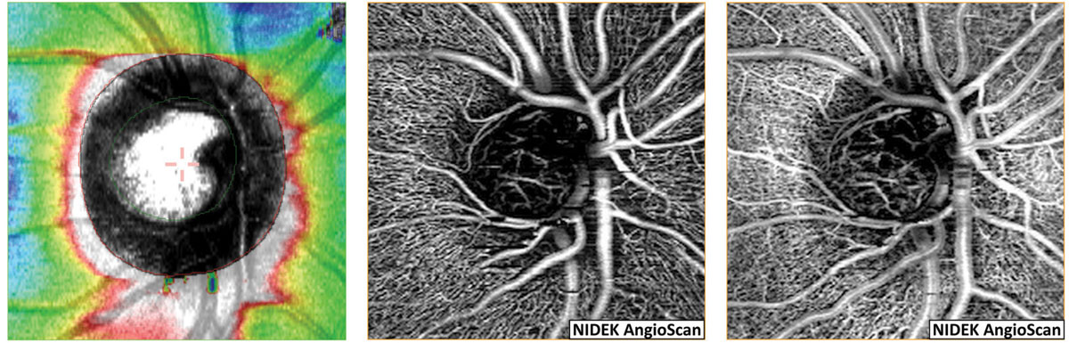 |
| Early OCT-A progression rate was found to be a slightly better predictor of future VF loss than early change rates in OCT RNFL or ganglion cell complex thickness. Photo: Carolyn Majcher, OD. Click image to enlarge. |
In this cohort, OCT-A imaging showed 83 eyes to be slow progressors and 84 eyes to be fast, with mean capillary density loss of -0.45% per year and -1.17% per year, respectively (mean difference -0.72%/year). Similarly, 83 eyes were slow OCT progressors, while 84 eyes were fast, with mean retinal nerve fiber layer thinning of -0.09μm per year and -0.60μm per year, respectively (mean difference -0.51μm/year).
The fast OCT-A and OCT progressors were associated with more rapid visual field loss (mean difference -0.18dB/year and -0.17dB/year, respectively). Fast OCT-A progressing eyes were more likely to have visual field progression (hazard ratio: 1.96). Also, 17 of 52 eyes (32.7%) with fast OCT-A and OCT progression developed subsequent visual field likely progression.
“Both trend and event analysis findings support the consideration of potential clinical use of OCT-A along with OCT for estimating the risk of glaucoma progression,” the researchers wrote in their paper.
The team did note that their results were from the initial three OCT or OCT-A visits instead of relying solely on one baseline test. “Although opting for more frequent testing may increase the predictability of visual field progression, such a testing paradigm may not be feasible in clinical practice,” they added.1
A commentary for the study also published in JAMA Ophthalmology highlighted that the fact that the findings were “synergistic with faster structural change in predicting faster future VF loss makes early OCT-A rate of change an important new biomarker of future VF progression.”2
The authors observed that “OCT-A may be useful in measuring change in eyes in which OCT has already reached the floor effect. Objectively and quantitatively identifying and predicting change in such eyes with advanced glaucoma could have great value.”
Still, the commentary emphasized that the study was unable to assess the relative benefit of OCT-A in advanced glaucoma, as the participants in this study had relatively mild damage; only 28 eyes with moderate to severe disease were included.
“It would be valuable in future work to understand the spatial association of RNFL thinning and decreasing ONH capillary density with the corresponding areas of future VF change,” the commentary authors wrote. “Novel parameters and further applications of OCT technology will allow clinicians to quantitatively identify and appropriately intervene for fast progressors who are at increased risk of visual loss and blindness.”2
1. Tansuebchuesai N, Nishida T, Moghimi S, et al. Rate of initial optic nerve head capillary density loss and risk of visual field progression. JAMA Ophthalmol. May 2, 2024. [Epub ahead of print]. 2. Schuman JS, Desai A, Lavinsky F. Optical coherence tomography angiography to predict visual field loss. JAMA Ophthalmol. May 2, 2024. [Epub ahead of print]. |
