 |
|
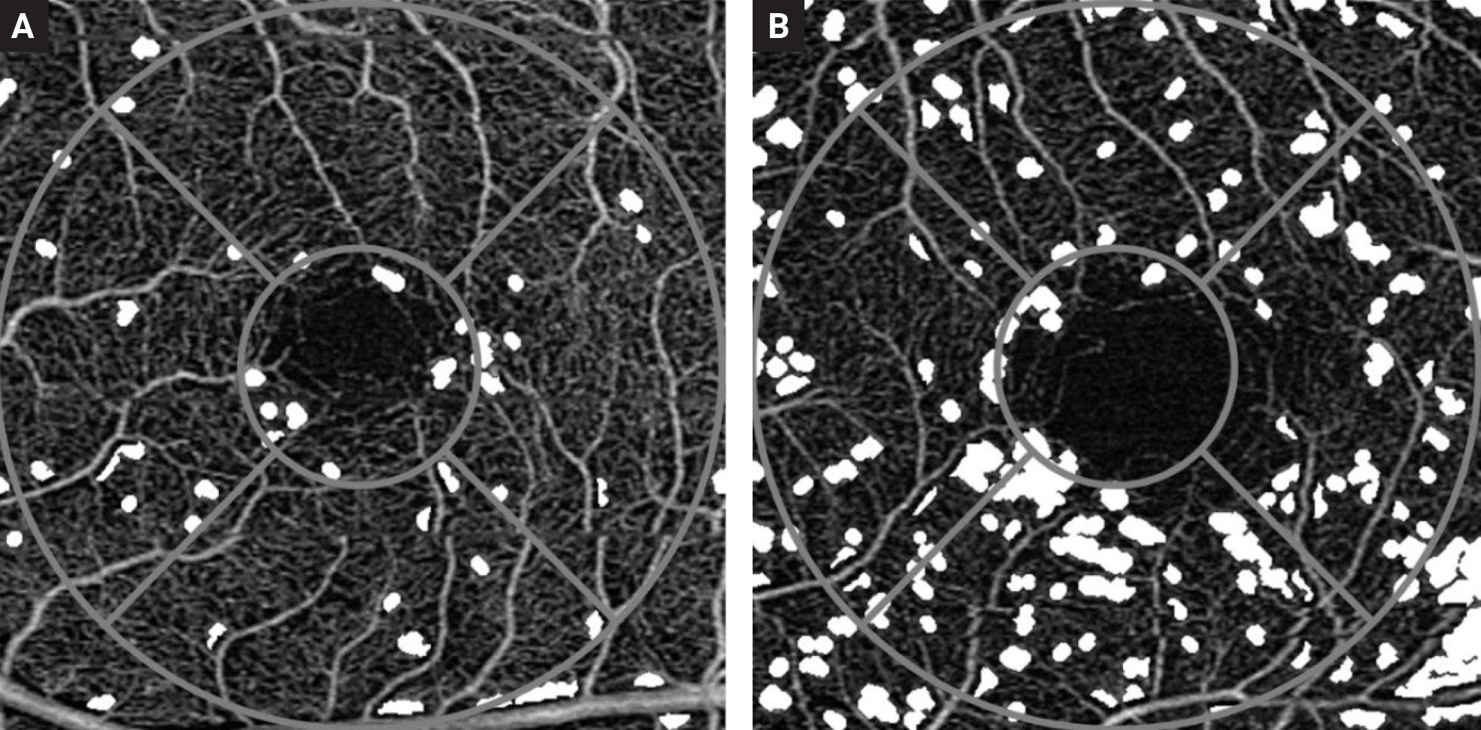
Nonperfused areas in the superficial capillary plexus are seen as white spaces in the intercapillary areas. Figure A is a 70-year-old subject without diabetes for comparison and figure B is the OCT-A scan of a 68-year-old diabetes patient without otherwise visible retinal lesions on fundus photography. Photo: Santos T, et al. Ophthalm Res. Oct. 11, 2023. Click image to enlarge. |
Identifying diabetic retinopathy (DR) early is of great importance, especially since not all patients have signs of the disease and very few present signs of vision-threatening retinopathy, a cause of blindness. It’s been shown that OCT-A and OCT can detect different stages of DR progression, so in a new study conducted in Portugal, researchers aimed to identify retinal microvascular changes using OCT-A in type 2 diabetes sufferers. They used the technology to identify vessel density and areas of capillary nonperfusion in the initial stages of DR, allowing for the identification of its ischemic phenotype very early in the disease process.
A total of 193 eyes of individuals with type 2 diabetes with no visible lesions in the fundus and identified after undergoing UWF-FP 200° exams were included.
Vessel density, perfusion density and areas of capillary nonperfusion values on OCT-A were significantly decreased when compared with healthy population. Density and nonperfusion values of the superficial capillary plexus (SCP) were more frequently decreased (35% and 45%, respectively) than density values of the deep capillary plexus (DCP) (9% and 15%), demonstrating that diabetic microvascular changes occur earlier in the SCP than in the DCP. The ischemic phenotype, identified by a definite decrease in vessel density or capillary nonperfusion in the SCP may, therefore, be identified in the preclinical stage of diabetic retinal disease, the researchers speculated.
The study shows that retinal capillary nonperfusion was detected by OCT-A—specifically metrics of vessel density and capillary nonperfusion —and can be identified in the central retina in eyes with type 2 diabetes before development of visible lesions in the retina. The microvascular changes lead to progressive increase in the areas of retinal nonperfusion which characterize the ischemic phenotype, associated with progression to the major vision threatening complications of diabetes, the authors explained. Because of this, retinal capillary nonperfusion of the SCP in preclinical retinopathy may be the biomarker that identifies the eyes at risk of progression to clinical retinopathy and potential vision loss.
“It is also particularly relevant that retinal capillary nonperfusion is associated with photoreceptor damage, identified by thinning of the OS retinal layer and presence of neurodegenerative changes demonstrated by thinning of the ganglion cell layer and inner plexiform layers,” the authors explained in their article for the journal Ophthalmic Research. “These observations indicate an early alteration of the neurovascular coupling unit in diabetic retinal disease and suggest an initial toxic neurodegenerative response to the chronic hyperglycemia of diabetes. Previous studies have documented subtle changes in color perception and contrast sensitivity in diabetic eyes without clinical retinopathy.”
The researchers; findings confirm the relevance of OCT-A to identify macular microvascular changes in the initial stages of DR, they argue, and suggest that the associations may be relevant for improved management of diabetic retinal disease, as achieving glucose control in the early stage could be the first evidence of retinal capillary nonperfusion in central retina—occurring before any detectable pathology.
“Furthermore, monitoring the rate of progression of retinal capillary nonperfusion may be the most appropriate monitor of diabetic retinopathy progression and development of vision-threatening complications,” they concluded.
Santos T, Santos AR, Almeida AC, et al. Retinal capillary nonperfusion in preclinical diabetic retinopathy. Ophthalm Res. Oct. 11, 2023. [Epub ahead of print.] |
