 |
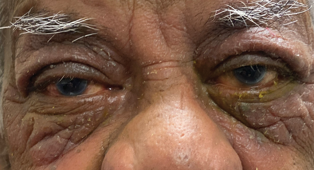
A 77-year-old Black man presented to the office with a chief complaint of “crusty eyes” of two weeks’ duration. Her vision was not affected but he said his eyes were “irritated.” He denied exposure to toxic chemicals but did report he was recently at an outdoor family function. Systemic and ocular history were unremarkable. He denied allergies of any kind.
Best-corrected entering visual acuities were 20/25 OD and 20/25 OS at distance and near. External examination is demonstrated in the photograph. There was no afferent pupil defect. Goldmann applanation tonometry measured 15mm Hg OU. The dilated fundus findings were normal peripherally and centrally with normal nerves and maculae.
The workup included photodocumentation and sensitivity testing. Additional questioning included hypersensitivity history to outdoor vegetation and/or animal dander and exposure.
 |
|
Is there anything visible in this photo from the exam that would suggest a possible diagnosis? Click image to enlarge. |
Skin Reaction
The diagnosis in this issue is contact dermatitis OU. The condition is an inflammatory reaction of the eyelid(s) and/or periocular adnexa. The presentation may occur unilaterally or bilaterally, depending upon the exposure. In acute cases, the patient may present with generalized lid erythema and edema, as well as pruritic vesicles or bullae.1 Chronic cases may show eczema, a characteristic thickening and scaling of the involved skin, which is typically seen in atopic dermatitis.2
Common symptoms in both the acute and chronic forms of contact dermatitis include intense itching and burning of the skin and eyes and associated tearing. The conjunctiva may also be involved, demonstrating variable bulbar injection and chemosis when included. Palpebral follicles may be seen in severe cases. Corneal involvement is rare, though chronic eye rubbing may lead to punctate keratopathy or epithelial erosion. Vision is not typically affected to any substantial degree and preauricular lymphadenopathy is characteristically absent.
Besides the hallmark “leathery” appearance of the skin, the history is important in correctly diagnosing contact dermatitis. Many cosmetics, cleaning agents, fabrics, fragrances, medications, nickel and even metallic jewelry can be implicated.2-4 Exposure to animal fur, dander or various plants (e.g., poison ivy) are additional vectors. Since the reaction connotes some acute exposure to an offending allergen, when the issue results from exposure in the home it is assumed the patient recently encountered “something “new” like a new perfume or laundry detergent. Many agents implicated in contact dermatitis represent weak sensitizers with exposures occurring over weeks, months or even years.4
In cases involving the ocular tissues, substances which are readily applied or coincidently touch the skin around the eyes include makeup (eyeliner, eye shadow, mascara) or makeup remover, sunscreen, contact lens solutions or metal spectacle frames. A wide range of ophthalmic medications may also have the capacity to induce allergic contact dermatitis.5
Subtypes
The condition may be subdivided into several categories: irritant contact dermatitis, allergic contact dermatitis and photoallergic contact dermatitis.
Irritant contact dermatitis (ICD) accounts for nearly 80% of all cases of contact dermatitis seen clinically.1 The condition, also referred to as non-allergic contact dermatitis, represents a non-preprogrammed, non-immunologic, local inflammatory reaction to a chemical or physical irritant.1 This may include substances such as soaps or detergents, solvents (e.g., paint thinner, acetone) or particulate matter such as fiberglass insulation.1
Typically, the severity of an ICD reaction is proportionate to the amount and duration of irritant exposure. The mechanism of action involves a direct, local cytotoxic effect on the epidermis, leading to subsequent keratinocyte damage.6 An ICD reaction can affect any individual, regardless of their immune status. A well-known example of ICD involves exposure to the plant Toxicodendron radicans, better known as poison ivy. Atopic individuals are especially susceptible to ICD and may have a lower threshold for irritant exposure.1,6
Allergic contact dermatitis (ACD), by its very definition, affects only individuals who are genetically “preprogrammed” toward allergic hypersensitivity to those particles. Like all allergic reactions, ACD occurs in two phases: a sensitization process, in which specific antibodies are generated toward the allergen, and an elicitation phase in which the actual cellular response occurs. The mechanism of ACD involves a delayed, or Type IV, hypersensitivity reaction.6 This is the cell-mediated response employing discrete subpopulations of T-lymphocytes and immunoregulatory cytokines, including tumor necrosis factor alpha and interleukins 1, 13 and 18.7 Mast cells, which play a pivotal role in immediate Type I hypersensitivity reactions such as allergic conjunctivitis, are not central to Type IV reactions. Mast cells still have an important function as they indirectly control neutrophil recruitment.8
Photoallergic contact dermatitis (PACD) is an eczematous skin reaction initiated by an otherwise benign substance on the skin that becomes noxious when exposed to ultraviolet light. We refer to substances that induce PACD as photosensitizing agents. PACD characteristically occurs only with areas of exposed skin, such as the hands and face. Known photosensitizing agents include tar-based products, octyl dimethyl PABA (an ingredient in some commercial sunscreen formulas), certain forms of vegetation (including carrot, lemon and mustard plants) some oral antibiotics, such as tetracycline, and topical NSAID medications.9,10
Management Approach
In treating contact dermatitis, attempts should be made to identify the causative agent. Often, this can be accomplished with a careful history but, in more challenging cases, epicutaneous patch testing can help to isolate the offending substance.11,12 Patch testing involves standardized samples of known allergens, placed on small delivery vehicles and applied to the skin of the upper back for two days.11,12 The test is typically performed only by an experienced dermatologist.
The ideal long-term solution is avoidance of the causative agent. Short-term management involves emollients, treatment of secondary infection (if present) and downregulation of the immune response. The easiest method of arresting and reversing the release of chemical modulators is the cold compress. Here, vasoconstriction slows the spread of edema and begins to limit the reaction-it is also immediately soothing.
Topical corticosteroids are the first-line medicinal therapy for most individuals, since these agents directly suppress the recruitment of polymorphonuclear leucocytes and reverse capillary permeability.12 Low-potency steroids such as hydrocortisone and desonide may be safer for use on the face, though stronger steroids such as triamcinolone (Aristocort) can be employed for moderate to severe disease.13 Steroid creams or lotions used twice daily for 10 to 14 days are usually very effective. Care must be taken to apply these preparations only to the skin of the lids and ocular adnexa, as they are not appropriate for use in the eye. Also, it is important to note that topical steroids may in some cases themselves be allergenic, confounding the management of these patients.14
Calcineurin inhibitors such as tacrolimus (Protopic ointment, Astellas Pharma) may benefit subjects who are poor candidates for topical corticosteroid therapy.12
More severe cases may require systemic corticosteroids such as oral prednisone (0.5 to 1mg/kg/d for two to three days, and tapered over one to two weeks) or a seven to 10-day steroid dose pak.1,6 The use of antihistamines is somewhat controversial in managing contact dermatitis.
Since histamine release from mast cells is not central to the pathophysiology of the disorder, antihistamines would seem to be superfluous. However, oral agents such as cetirizine 10mg (over-the-counter Zyrtec) or desloratadine 5mg (over-the-counter Clarinex) once daily may help to curtail itching and hence may be beneficial as an addition to topical corticosteroid therapy.
For any associated conjunctivitis topical local supportive therapy is beneficial. This includes first and foremost cold compresses and liberal use of ocular lubricants. Topical over-the-counter antihistamine/mast cell stabilizer products (e.g., Pataday, Lastacaft or Alaway once daily) may provide palliative relief of conjunctival itching, as well as ocular swelling and hyperemia. More severe involvement, however, may warrant topical ophthalmic corticosteroids such as 1% prednisolone acetate or 0.5% loteprednol etabonate every two to four hours for several days.
Takeaways
In general, the skin of the eyelid, by virtue of its anatomically thin structure, is particularly susceptible to inflammation by irritant or allergic contact dermatitis. Profound reactions may be seen in this area. The differential diagnosis of contact dermatitis must include more extreme conditions such as preseptal cellulitis, orbital cellulitis, chlamydial infection, oculoglandular conjunctivitis and carotid cavernous fistula. A vesicular or pustular presentation warrants investigation for herpes zoster or simplex blepharitis. This can be ruled out by assessing the region for facial excoriations, increased temperature and pain-to-the-touch.
Contact dermatitis is self-limiting in the case where the inciting agent is identified and removed. Patients should be counseled that topical and oral steroids can raise intraocular pressure as well as thin the skin. Anyone placed on these agents must be reassessed to rule out these complications.
This patient was placed on an over-the-counter antihistamine (diphenhydramine) by mouth Q12h, cold compresses and over-the-counter cortisone 10mg anti-inflammatory cream BID. An over-the-counter topical mast cell stabilizer antihistamine eye drop was recommended for ocular symptoms. The condition resolved slowly over five days without complications.
Dr. Gurwood is a professor of clinical sciences at The Eye Institute of the Pennsylvania College of Optometry at Salus University. He is a co-chief of Primary Care Suite 3. He is attending medical staff in the department of ophthalmology at Albert Einstein Medical Center, Philadelphia. He has no financial interests to disclose.
1.Mark BJ, Slavin RG. Allergic contact dermatitis. Med Clin North Am. 2006;90(1):169-85. 2. Watkins S, Zippin J. Allergic contact dermatitis and cosmetics. Cutis. 2012;90(4):201-4. 3. Brandão MH, Gontijo B. Contact sensitivity to metals (chromium, cobalt and nickel) in childhood. An Bras Dermatol. 2012;87(2):269-76. 4. Biebl KA, Warshaw EM. Allergic contact dermatitis to cosmetics. Dermatol Clin. 2006;24(2):215-32, vii. 5. Mughal AA, Kalavala M. Contact dermatitis to ophthalmic solutions. Clin Exp Dermatol. 2012;37(6):593-7; quiz 597-8. 6. Usatine RP, Riojas M. Diagnosis and management of contact dermatitis. Am Fam Physician. 2010;82(3):249-55. 7. Incorvaia C, Frati F, Verna N, et al. Allergy and the skin. Clin Exp Immunol. 2008;153 Suppl 1:27-9. 8. Biedermann T, Kneilling M, Mailhammer R, et al. Mast cells control neutrophil recruitment during T cell-mediated delayed-type hypersensitivity reactions through tumor necrosis factor and macrophage inflammatory protein 2. J Exp Med. 2000;192(10):1441-52. 9. Deleo VA. Photocontact dermatitis. Dermatol Ther. 2004;17(4):279-88. 10. Kerr A, Ferguson J. Photoallergic contact dermatitis. Photodermatol Photoimmunol Photomed. 2010;26(2):56-65. 11. White JM. Patch testing: what allergists should know. Clin Exp Allergy. 2012;42(2):180-5. 12. Morris S, Barlow R, Selva D, Malhotra R. Allergic contact dermatitis: a case series and review for the ophthalmologist. Br J Ophthalmol. 2011;95(7):903-8. 13. Li LY, Cruz PD Jr. Allergic contact dermatitis: pathophysiology applied to future therapy. Dermatol Ther. 2004;17(3):219-23. 14. Gonul M, Gul U. Detection of contact hypersensitivity to corticosteroids in allergic contact dermatitis patients who do not respond to topical corticosteroids. Contact Dermatitis. 2005;53(2):67-70. |
