 |
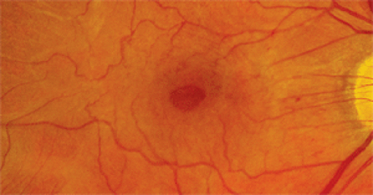
| Tractional forces from epiretinal membrane or vitreous adhesion were identified in patients in any lamellar macular hole stage. Mark Dunbar, OD. Click image to enlarge. |
Although the idiopathic lamellar macular hole has been investigated extensively, only a few studies have examined the structural changes both before and after formation. The development and evolution of lamellar macular holes in highly myopic eyes may be more complicated, which is what prompted researchers of a recent study to evaluate the development, evolution, outcomes and prognostic factors in affected patients.
Pre- and post-lamellar macular hole OCT findings and visual acuity were collected from 50 eyes of 47 highly myopic patients. Structural progression was defined as an increase in the height of retinoschisis and the development of foveal detachment, full-thickness macular hole or retinal detachment.
Four types of lamellar macular hole developmental processes were observed. Type one started from foveal tissue avulsion caused by abnormal posterior vitreous detachment and mimicked the abortive development of full-thickness macular holes. Types two and three originated from ruptured parafoveal cysts/schisis and central foveal cysts, respectively. Type four was induced by the persistent epiretinal membrane traction causing progressive central foveal thinning without going through the parafoveal or foveal cyst stages.
In the pre- and early lamellar macular hole stages, tractional forces from epiretinal membrane or vitreous adhesion could be identified in all cases, suggesting that traction, instead of degeneration, was the primary mechanism of lamellar macular hole formation in highly myopic eyes.
“Lamellar macular holes in highly myopic eyes formed by epiretinal membrane induced progressive foveal thinning in the absence of an intervening stage of cyst or retinoschisis and tended to progress into a full-thickness macular hole with the worst visual outcome,” the authors explained. “Lamellar macular holes with retinoschisis were more susceptible to anatomical progression than simple lamellar macular holes in highly myopic eyes. Lamellar macular holes may develop before, after or simultaneously with the retinoschisis.”
The study concluded that lamellar macular holes in highly myopic eyes may develop through different formation processes, and all of them involve traction. Lamellar macular holes with foveal thinning induced by epiretinal membranes had the worst outcomes, and degenerative configuration may occur in some stages during the evolution process.
Hsia Y, Lee CY, Ho TC, et al. The development and evolution of lamellar macular hole in highly myopic eyes. Eye. May 13, 2022. [Epub ahead of print]. |
