 |
|
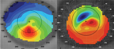
Keratoconus patients who don't wear contacts are likely to experience a similar level of endothelial cell changes regardless of disease stage. Click image to enlarge. |
Understanding how endothelial changes may present in different stages of keratoconus is critical in choosing an appropriate treatment or surgical plan for your patient. While studies observing keratoconus-related changes in this layer of the cornea have been minimal, researchers from a hospital in India recently set out to change that. They used a specular microscope to compare various parameters across keratoconus stages in a group of non-contact wearing patients, finding that very few changes are actually associated with keratoconus severity.
The researchers included 162 eyes of 96 patients with keratoconus in one or both eyes who visited their practicing hospital between April 2018 and December 2019. Each patient underwent a detailed ophthalmic examination. Endothelial cell changes were closely observed with an SP-1P specular microscope, a newer generation capable of capturing various measurements such as endothelial cell density, coefficient of variation, frequency of hexagonal cells, number of cells analyzed and the minimum, maximum and average cell area.
The analysis revealed very few differences in endothelial cell parameters between the four stages of keratoconus under investigation. Changes in the following parameters were not correlated with keratoconus severity: endothelial cell density, coefficient of variation, frequency of hexagonal cells and minimum, maximum and average cell area between the stages. However, it’s important to note that measurement was not possible in eyes with stage four keratoconus.
The only variable parameter was the number of analyzed cells, which the researchers found decreased with severity of keratoconus, despite never falling below 100. Still, this parameter demonstrated a weak spearman rank correlation. The researchers noted, “A possible explanation for this decrease in number could be because of the increase in protrusion and stretching of the cornea as the keratoconus stage progresses.”
In conclusion, these findings show that the severity of keratoconus does not affect the corneal endothelium between stages zero to three in non-contact lens wearers. In future studies, the endothelial changes in more corneal zones should be assessed using a modern specular microscope, which can also capture data from the paracentral and midperipheral cornea, as opposed to only the central cornea.
Shah Z, Shilpy N, Purohit D. Assessment and correlation of corneal endothelial cell changes in different stages of keratoconus in non-contact lens wearers. Optometry Vis Sci. September 9, 2021. [Epub ahead of print]. |
