 |
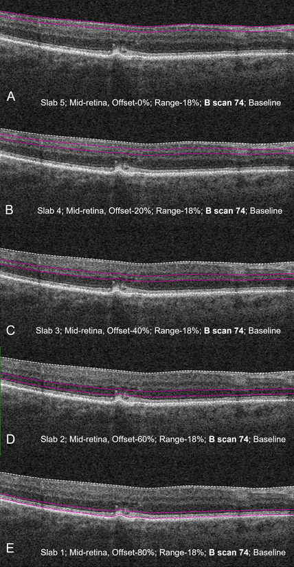
Structural OCT B-scan with corresponding slab locations showing the presence of intra-retinal hyperreflective foci. These lesions in the inner and outer retina may have disparate origins and pathophysiology, as evidenced by the inequal abundance in the outer retina. Photo: Sadda S, et al. Res Sq. Sept. 20, 2023/Creative Commons 4.0. Click image to enlarge. |
OCT has been able to identify major biomarkers for many diseases. More recently, it has accounted for one biomarker of development to late-stage age-related macular degeneration (AMD). The presence and distribution of intraretinal hyper-reflective foci (IHRF) in individuals with intermediate stage AMD has been linked to greater chance of progression. Building on this foundation, researchers of one recent study assessed the change in number and distribution of IHRF over two years.
Axial distribution of IHRF was quantified in intermediate AMD eyes at baseline and 24 months, conducted with a series of five sequential, equidistant en face OCT retinal slabs generated between the internal limiting membrane’s outer border and retinal pigment epithelium’s (RPE) inner border.
Of the 1,339 individuals included at baseline, 52 eyes displayed evidence of IHRF, all continuing to show evidence at the 24-month follow-up. Total average IHRF count/eye increased from baseline and at 24 months, 76.9% of eyes showed IHRF increase, 15.4% showed decreases and 7.6% showed no change. On average, IHRF increase occurred at a rate of 2.5x fold. The outer slabs displayed greater numbers and greater increase in IHRF at the 24-month mark.
Despite a majority displaying increased IHRF, presence can vary from lesions appearing or disappearing at a specific site. The most dramatic IHRF count expansion occurred in the outermost retinal layers; counts for inner layers were more stable.
More specifically, the study authors note that IHRF counts increased in about two-thirds of cases in outer slabs 1 and 2. Only 11.5% of cases with increases occurred in slab 4 and no IHRF appeared in the innermost slab 5. This finding contradicts previously reported progressive inward IHRF migration.
The study’s results do suggest, however, that a disparate increase in IHRF occurs over time in the outer retina up to the deep capillary vascular plexus (DCP) but does not continue beyond this level. The researchers believe this to be due to RPE cells’ inward migration triggered by underlying choriocapillaris ischemia, with moving inward presenting possible perfusion and nourishment from the retinal DCP. Once this area is reached, no further ischemic drive or oxygenation is there to pull the cells further. The lower incidence of IHRF lesions in inner retinal levels, the authors surmise, may be from other inflammatory or microglial sources.
Finally, the authors point to the dramatic increase seen generally in IHRF count over the two years. As they articulate, “this suggests that the appearance on IHRF may represent a critical tipping point in the eye, highlighting an eye that might be in severe distress. Once an IHRF appears, there may be dramatic acceleration with many more IHRF appearing. This is not surprising, as IHRF are perhaps the strongest biomarker for progression to late AMD and atrophy.”
Fortunately, some analyses suggest therapeutic agents administered earlier in the disease process may be more effective, thus reducing conversion from incomplete RPE and outer retinal atrophy to the complete version. From this, the authors posit that “earlier intervention at the first appearance of IHRF may be a target for future therapies and trials and prevention of an increase in IHRF could potentially serve as an endpoint to access effectiveness of treatment.”
Sadda S, Verma A, Corradetti G, et al. Longitudinal evaluation of the distribution of intraretinal hyper-reflective foci in eyes with intermediate age-related macular degeneration. Res Sq [Preprint]. September 20, 2023. [Epub ahead of print]. |
