 |
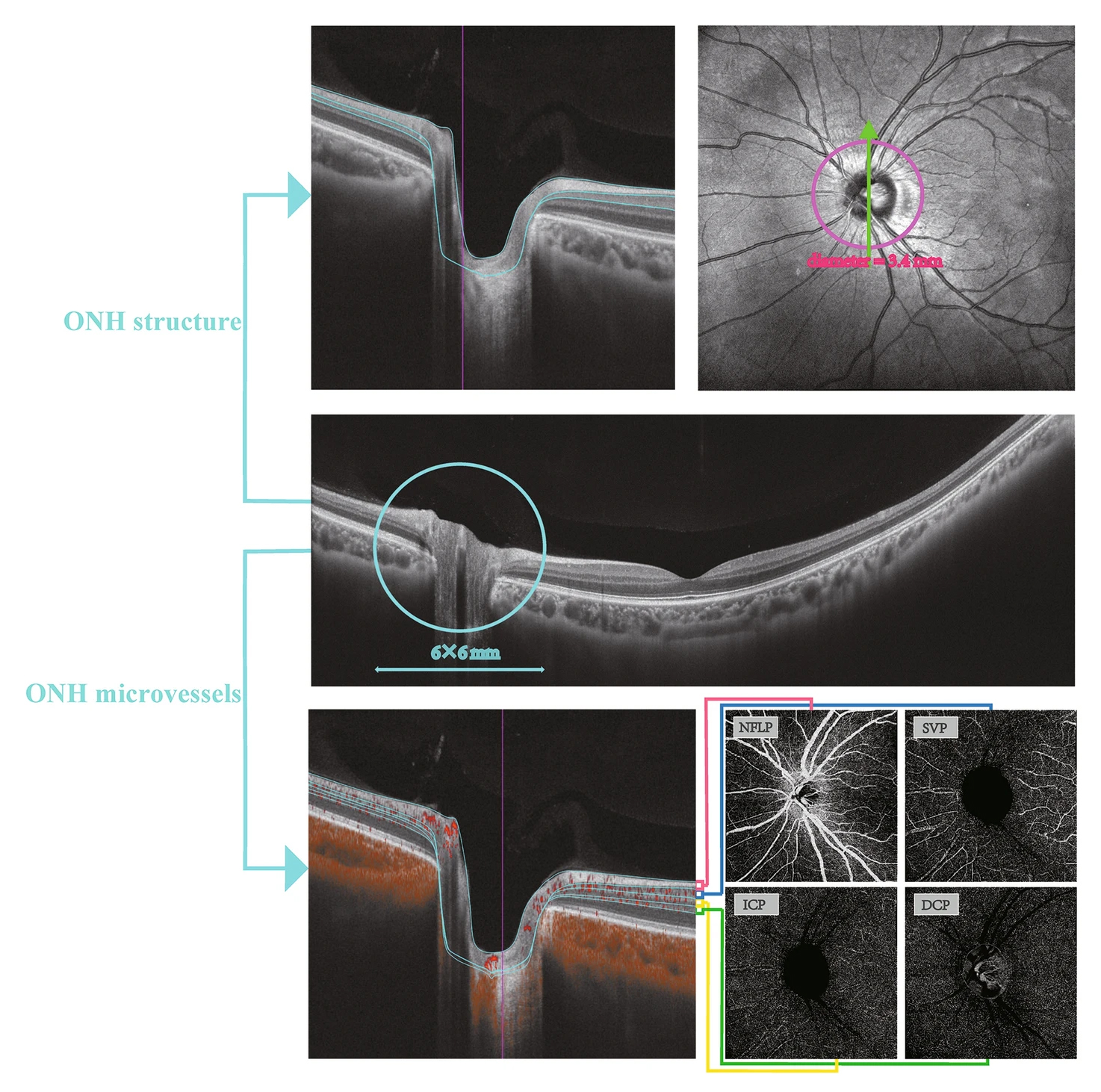
| Objective clinical tools such as OCT and OCT-A can offer reliable data on visual acuity changes, such as macular structural measurements. Photo: Wang H, et al. Ophthalmol Ther 2023. Click image to enlarge. |
Monitoring for papilledema development in patients with increased intracranial hypertension is critical, as this finding is a strong indicator of visual loss and impairment and may lead to optic nerve atrophy. Currently, experts say improvement in reliable and sensitive monitoring is needed. One possible option is using swept-source OCT and OCT angiography. Researchers in China used these modalities to study optic disc morphology and several structural and microvascular parameters. According to their study, published recently in Ophthalmology and Therapy, patients with intracranial hypertension exhibit vascular abnormalities that correlate with clinical manifestations of the condition.
The researchers found that patients with intracranial hypertension (n=61) had significantly thicker peripapillary retinal nerve fiber layer and ganglion cell inner plexiform layer thickness with larger optic nerve head rim area than controls (n=65). Additionally, they observed significantly increased microvascular densities in the nerve fiber layer plexus and significantly reduced densities in the superficial vascular plexus, intermediate capillary plexus and deep capillary plexus vs. controls.
Importantly, the researchers reported that structural thickness and microvascular densities correlated significantly with Frisen scores and intracranial pressure in patients with intracranial hypertension. This highlights “the potential clinical relevance of these parameters,” the researchers wrote in their paper.
They concluded that “patients with intracranial hypertension may benefit from monitoring optic nerve head structure and microvascular changes” with the OCT/OCT-A technology.
Wang H, Cao L, Kwapong WR, et al. Optic nerve head changes measured by swept source optical coherence tomography and angiography in patients with intracranial hypertension. Ophthalmol Ther. October 4, 2023. [Epub ahead of print.] |
