 |
| Several small studies have suggested children with FASD may be at an increased risk of various refractive and ocular conditions. A more recent, large study identified strabismus as a potential visual outcome of the disorder, but only in those with partial fetal alcohol syndrome. Photo: Andrew Gurwood. Click image to enlarge. |
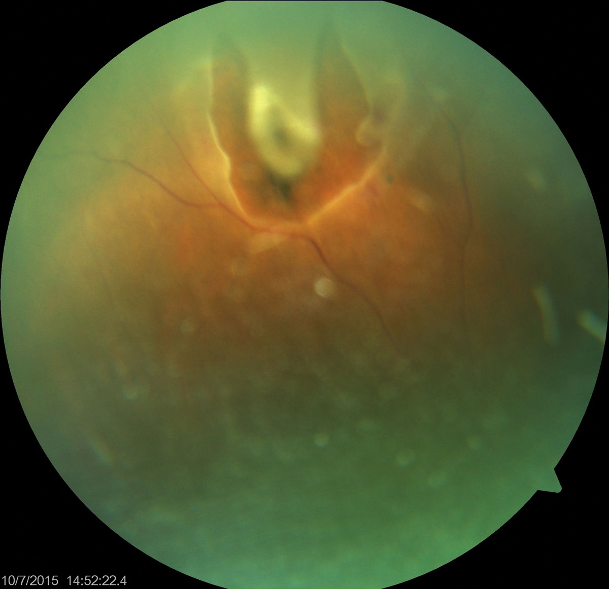
In certain telemedicine situations, imaging can make up for the lack of an in-person evaluation but not when it comes to detecting horseshoe tears. According to a paper published recently in the American Journal of Ophthalmology, which assessed the detection sensitivity of ultra-widefield imaging in isolation (as opposed to the typical combined approach with scleral depression), this approach on its own missed about half of horseshoe tears.
The retrospective analysis (123 patients; 135 horseshoe tears) showed that 51.1% of horseshoe tears were visualized using ultra-widefield images and 48.9% were not. The researchers reported the following sensitivities for identifying horseshoe tears:
- 17.1% for the superior quadrant
- 32% for the inferior quadrant
- 50% for the nasal quadrant
- 85.5% for the temporal quadrant
Ultra-widefield imaging performed poorly in the superior and inferior quadrants. The researchers explained in their paper that low sensitivity in the superior quadrant in particular is a cause for concern since missed horseshoe tears in this region are at a greater risk for progression to retinal detachment.
“Considering the urgency of treating horseshoe tears and the potential for significant visual morbidity from retinal detachments, it’s crucial to highlight that ultra-widefield imaging shouldn’t be relied upon as a substitute for funduscopic examination in the evaluation of posterior vitreous detachment or symptoms indicative of a horseshoe tear,” the researchers concluded in their AJO paper. “This cautionary note is essential as a negative ultra-widefield image can provide false reassurance, potentially leading to delayed diagnosis and management of a critical retinal condition.”
Lin AC, Kalaw FG, Schönbach EM, et al. The sensitivity of ultra-widefield fundus photography versus scleral depressed examination for detection of retinal horseshoe tears. Am J Ophthalmol 2023. [Epub ahead of print]. |
