 |
|
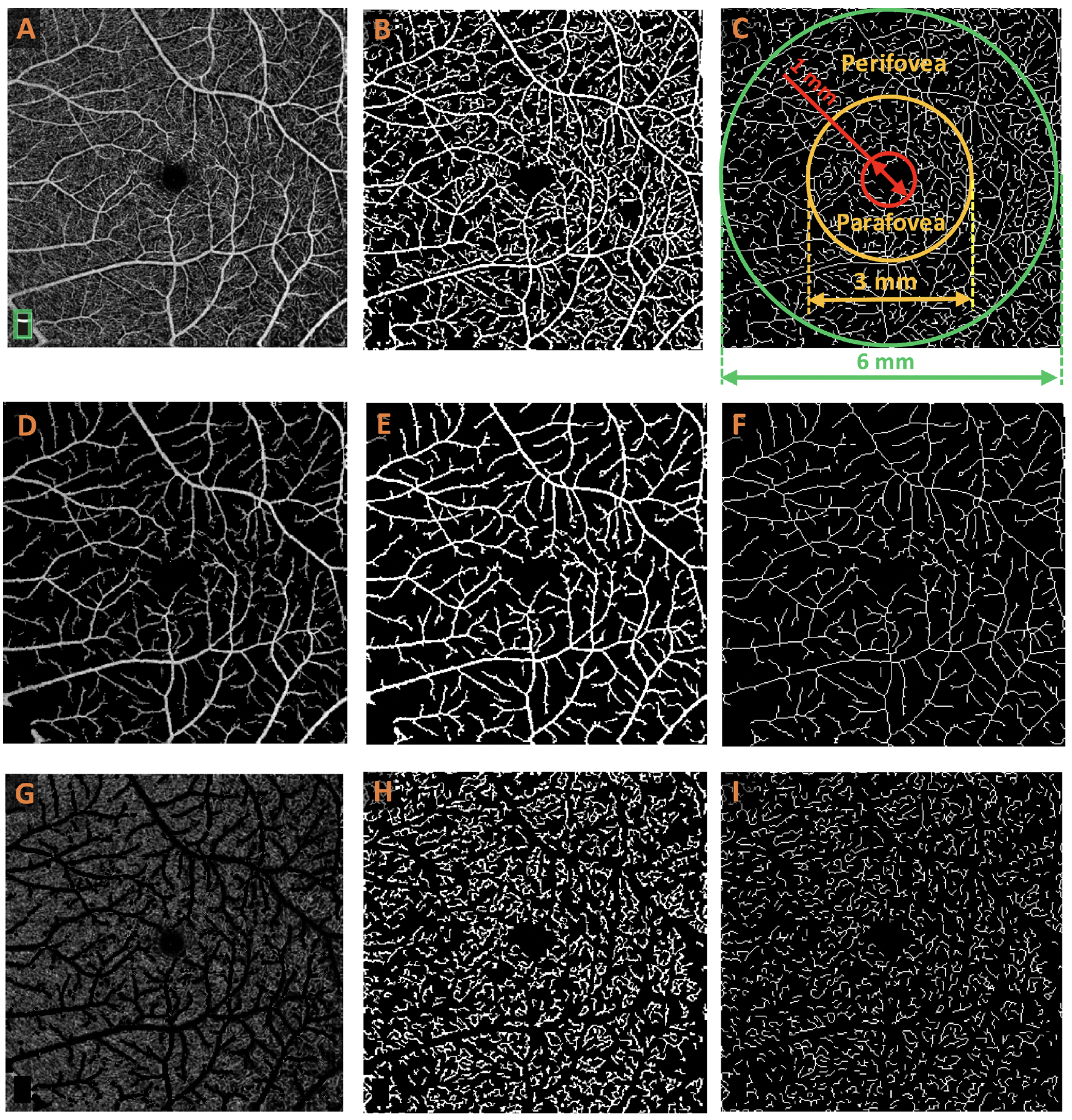
Using a new OCT-A technique to isolate capillaries improved DR classification performance. These images from the study show the process of feature extraction. An OCT-A image of a total vasculature map (A) is captured and then binarized (B) to isolate the vasculature, and then skeletonized (C) to illustrate fovea, parafovea and perifovea areas. Then, a large vessel map (D) is extracted, binarized (E) and skeletonized (F). Next, a capillary map (G) is produced, binarized (H) and finally skeletonized (I). Photo:Abtahi M, et al. Invest Ophthalmol Vis Sci. 2024;65(10):20. Click image to enlarge. |
Diabetic retinopathy (DR) manifests primarily in microvasculature. It characteristically presents capillary damage, seen clinically in findings like microaneurysms, hemorrhages and cotton-wool spots. However, as the disease progresses, it may also affect larger vessels, leading to symptoms of beading and potential vascular occlusion. In fact, in advanced DR, OCT angiography (OCT-A) is able to detect venous beading and intraretinal microvascular abnormalities.
Consequently, researchers from the University of Chicago recently published on how to use a new OCT-A technique called differential capillary–large vessel (CLV) analysis to differentiate between the two structures in evaluating DR or other possible retinal conditions that could impact capillaries. Integration of CLV analysis into clinical imaging devices like OCT-A would allow for the possibility of improving disease detection and accuracy, the researchers argue, using a noninvasive method that can help identify microvascular distortions with capillary-level resolution.
The team analyzed 212 OCT-A images from 146 patients. Of this cohort, 28 were controls, 36 were diabetic patients without DR, 31 had mild nonproliferative DR (NPDR), 28 had moderate NPDR and 23 had severe NPDR.
Differential CLV analysis was performed by extracting quantitative features from the entire image as well as the parafovea and perifovea regions. The analysis improved accuracy of DR determination, with binary classification (i.e., DR or no DR) accuracy improving by 11.8%—rising from 77.5% to 89.3%. With multiclass classifications (i.e., which stage of DR), accuracy increased by 7.6% from 78.7% to 86.2%. Binary classification accuracy improved even further when incorporating features from the entire image and both para- and perifovea areas, from 83.1% to 93.8%, while multiclass accuracy using these combined features raised from 82.6% to 87.9%.
In the discussion section of their paper for Translational Vision Science & Technology, the study researchers elaborate on how they were able to analyze these features, stating that, to their knowledge, this is the first study that applied differential CLV analysis in OCT-A for DR classification. The novel technique segregated capillaries and large vessels within OCT-A images—facilitating comprehensive CLV analysis. Five OCT-A features were computed—perfusion intensity density, blood vessel density, vessel skeleton density, blood vessel caliber and vessel area flux—for total vasculature, large vessels and capillaries along with their corresponding ratios across varying regions. It was the ratios that allowed them to quantify and analyze the relative feature changes in total vasculature, large vessels and capillaries.
The authors further explain that DR classification using only features from the capillary network yielded very comparable results as with using all features. From this, they can extrapolate that “focusing on capillary features after differential CLV analysis could simplify and enhance the diagnostic process, underscoring the critical role of the capillary network in early DR detection,” they wrote.
More generally, their findings “provide valuable insights into the potential integration of CLV analysis into clinical applications, aiming to improve sensitivity for effective disease diagnosis, treatment and management,” the paper concludes.
Abtahi M, Le D, Ebrahimi B, et al. Differential capillary and large vessel analysis improves OCT-A classification of diabetic retinopathy. Invest Ophthalmol Vis Sci. 2024;65(10):20. |