 |
| En face OCT imaging can help with the detection of AMD, which was validated in this recent study. Photo: Carolyn Majcher, OD. Click image to enlarge. |
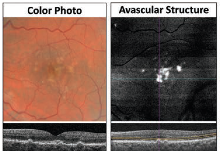
A recent retrospective analysis explored a number of different imaging modalities, including en face OCT, for the detection and monitoring of calcified drusen among patients with nonexudative age-related macular degeneration (AMD). The researchers performed a review of same-day color fundus, fundus autofluorescence, near infrared and en face swept-source OCT images. They used the different imaging modalities to compare the appearance and progression of calcified drusen in this patient population.
The analysis included 220 eyes from 139 nonexudative AMD patients. Of these, 42.7% contained calcified drusen either at baseline or during follow-up. Additionally, the data showed that six eyes had some calcified drusen regress without developing complete retinal pigment epithelium and outer retinal atrophy during follow-up, according to the study authors. This was confirmed across imaging modalities.
The researchers found that calcified drusen appeared as dark focal lesions, or choroidal hypotransmission defects, on the en face swept-source OCT images. They also reported that the corresponding B-scans revealed drusen with heterogeneous internal reflectivity, hyporeflective cores and hyperreflective caps.
“In the majority of the calcified drusen, choroidal hypertransmission defects were observed to develop over time around the periphery of the hypotransmission defects giving them the appearance of a donut lesion on the en face SS-OCT images,” the study authors explained. They noted that these donut lesions were correlated with significant attenuation of the overlying retina. Hypoautofluorescence was observed at the location of these lesions, according to the corresponding fundus autofluorescence images.
“Our results support previous findings showing that these prevalent lesions are an important risk factor for the progression of complete retinal pigment epithelium and outer retinal atrophy,” the study authors stated. “We also demonstrated that en face OCT imaging can detect calcified drusen through the presence of choroidal hypotransmission defects, which can progress to donut lesions and complete retinal pigment epithelium and outer retinal atrophy.”
They concluded that using en face OCT imaging for the identification and monitoring of calcified drusen could be a valuable tool when assessing the risk of disease progression among patients with AMD.
Liu J, Laiginhas R, Shen M, et al. Multimodal imaging and en face OCT detection of calcified drusen in eyes with age-related macular degeneration. Ophthalmol Sci. April 20, 2022. [Epub ahead of print]. |
