Q. Im finding that keratometry and A-scan ultrasonography do not always accurately determine the IOL correction on patients who have undergone refractive surgery. Why does this happen, and how can we assure a better outcome?

A. The choice of IOL is based partly on the patients corneal curvature. Because refractive surgery alters the curvature, its more difficult to accurately measure the cornea and calculate the correct IOL power.
Standard keratometers do not give accurate readings [on post-refractive surgery patients], says optometrist Howell Findley, director of Commonwealth Eye Surgery Center in Nicholasville, Ky. Therefore, special care must be given to obtain true keratometry readings for IOL power calculation formulas.
Realize that keratometers and A-scan ultrasonographers were designed before the era of refractive surgery. These instruments make assumptions about the anatomy and refractive properties of the cornea that are no longer valid following most keratorefractive procedures. These breakdowns in IOL calculation often result in a refractive surprise after cataract surgery, which may require surgical correction.1
However, there are steps you can take prior to surgery to help patients avoid this outcome.
Since accurate keratometry and A-scan measurements are the most important factors in IOL calculation, a number of methods have been proposed to adjust the biometry readings post-refractive surgery, says Fargo, N.D., optometrist Heather Cowden.
These include:
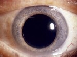
IOL correction on patients who have had refractive surgery can be difficult.
Clinical history method. There are many variations on this method, but it is generally considered the most accurate option.2 To use this method, however, youll need to know the patients keratometry readings before the refractive surgery. With this method, you calculate IOL power by subtracting the refractive change that occurred as a result of surgery from the initial (pre-operative) keratometry readings. For example, if the pre-op Ks were 45.00D and the patient went from 5.00D to plano, the patients true keratometry reading is now 40.00D.
This method highlights the importance of having a good set of Keratometry readings recorded in the chart of every patient you refer for refractive surgery.
Contact lens method. This method is useful if pre-operative keratometry readings are not available, Dr. Cowden says.
However, to use this method, patients should have best corrected acuity of at least 20/60.
Place a few drops of topical anesthetic on the patients eye, insert a plano gas permeable lens (with a known base curve) and perform an over-refraction. If the curve of the post-op eye matches that of the rigid lens, the spheroequivalent with the contact lens in place will be the same as without, says Dr. Findley.
To arrive at the true post-op corneal power using the contact lens method, subtract the spheroequivalent of the over-refraction from the known base curve of the contact lens. For instance: If the contact lens you use has a base curve of 42.00D and the over-re-fraction is 1.50D, the correct IOL power is 43.50 (42.00 minus -1.50).
One study comparing the efficacy of different methods to calculate IOL power found when corneal power is known prior to refractive surgery, the contact lens method produced the most accurate results.3
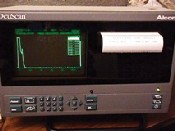
Average topography. Many corneal topographers have the ability to take keratometry readings at 3mm, 5mm and 9mm intervals, then provide a detailed map of the patients corneal curvature.
Dr. Findley says you should generally use the keratometry readings taken from the 3mm zone to calculate IOL power with this method. Axial length measuring devices incorporate an internal formula that calculates the IOL power.
To use this method, simply enter the patients keratometry readings (preferably from a topography unit) into the IOLmaster and the device will automatically calculate the IOL power for you.
Q. What is the O.D.s role in advising the surgeon about desired post-op refraction?
A. The optometrist has an important role to play in obtaining satisfactory patient outcomes, Dr. Findley says. As the comanaging doctor, you can:
Provide the surgeon with historical data on pre-op keratometry readings and post-op results. This information can be critical, especially if the cataract surgeon was not the refractive surgeon.
Accurate keratometry and A-scan measurements are key in IOL calculation.
Counsel the patient to have realistic expectations. Be sure your patients know that because they had refractive surgery, it is much more difficult to accurately pinpoint the IOL power.
Recommend whether monovision is appropriate for a given patient. Myopes and those who do not require good depth perception generally respond best to monovision correction. However, patients with prolonged daily near tasks may not be ideal candidates for monovision. Amblyopia, type-A personalities or insistence on a 20/20 surgical outcome should send up red flags, Dr. Cowden says.
If you think surgical monovision is an option for your patient, try a three-week trial using a silicone hydrogel contact lens, Dr. Cowden suggests. Then, be sure to communicate your patients preference to the surgeon, either by letter or telephone (or better yet, both), and well in advance of the surgery.
1. Hamilton DR, Hardten DR. Cataract surgery in patients with prior refractive surgery. Curr Opin Ophthalmol 2003 Feb;14(1):44-53.
2. Argento C, Cosentine MJ, Badoza D. Intraocular lens power calculation after refractive surgery. J Cataract Refract Surg 2003 Jul;29(7):1346-51.
3. Kim J, Lee D, Joo C. Measuring corneal power for intraocular lens power calculation after refractive surgery. Comparison of methods. J Cataract Refract Surg. 2002 Nov;28(11):1932-8.
