
Neuro-optometric cases can be intimidating and challenging due to their seemingly complex nature. However, optometrists can learn to correctly diagnose and comanage these patients by systematically learning to evaluate the patients demographics, signs, symptoms and associated diseases.
Here are four commonly encountered neuro-optometric cases in an easy-to-follow guide to pathophysiology and diagnosis.
Case 1: Unilateral Vision Loss And Optic Nerve Edema
History. Chuck is a 60-year-old white male who presents for a yearly exam. He reports a sudden, painless decrease in the visual acuity of his right eye for approximately two weeks. His systemic history is positive for hypertension and hyperlipidemia, which he says he controls with medication.
Diagnostic Data. His entering best-corrected visual acuity is 20/200 O.D. and 20/25 O.S. Initial testing finds an afferent pupillary defect (APD) in his right eye. All other preliminary testing is normal. The rest of the exam is normal, except for the presence of optic nerve edema in the right eye (figure 1). The left optic nerve is normal with 0.2 cupping (figure 2). The patient reports no other symptoms.
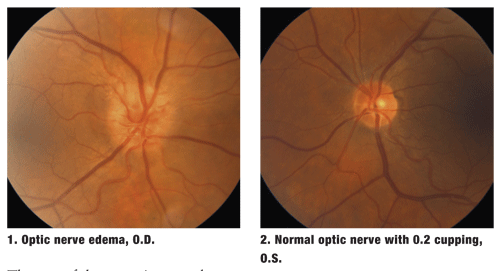
Diagnosis. In this case, we have an older patient with a painless, unilateral loss of vision with systemic vasculopathic conditions, an APD, a normal retinal exam, and a unilateral, abnormal optic nerve O.D. Unilateral, optic nerve swelling is seen in a variety of ocular disease states (see Common Conditions with Unilateral Optic Nerve Edema), but the patients normal retinal exam, systemic conditions, age, gender and race point to a likely diagnosis of non-arteritic ischemic optic neuropathy (NAION). In addition, our patient doesnt report any typical signs of arteritic disease (also known as giant cell arteritis), such as headache, jaw claudication, fatigue or scalp tenderness.
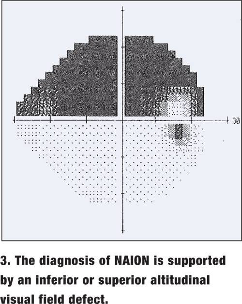 In NAION, visual acuity is mildly to seriously reduced. In the absence of other testing, the diagnosis is further supported by an inferior or superior altitudinal visual field defect (figure 3), although this is not pathognomonic.
Another clinical pearl when considering a diagnosis of NAION:
A small, crowded optic nerve head, also known as a disc-at-risk, is more likely to develop NAION than a larger cup.
In evaluating arteritic disease vs. non-arteritic disease, segmental optic nerve swelling and hemorrhages can help support a NAION diagnosis, while giant cell arteritis more characteristically has a pale optic nerve with cotton-wool spots.1
Treatment/Management. First, order an erythrocyte sedimentation rate (ESR), a complete blood count (CBC), and a C-reactive protein test to rule out arteritic disease, even without the associated symptoms. Order the bloodwork yourself, or refer the patient to an ophthalmologist or general practitioner. Because the arteritic form can progress rapidly, treat suspected cases as an ocular emergency due to the increased likelihood of bilateral involvement and the urgent need for systemic steroids and a temporal artery biopsy.2
For further evaluation, all patients with optic nerve swelling should undergo magnetic resonance imaging (MRI) to rule out intraorbital or intracranial disease. Also, if you suspect any untreated systemic diseases, refer the patient to the appropriate practitioner for further evaluation.
There are no definitive treatments for NAION. However, one-third of patients develop NAION in the fellow eye, so the best therapy is controlling the associated systemic conditions.3 The decreased visual acuity, associated field loss and APD are generally permanent, although mild improvements may be seen as the optic nerve edema resolves over four to eight weeks.4 Any optic nerve pallor is permanent.
Due to the associated vasculopathic conditions, the patient was instructed to follow-up with his general physician for further assessment of his hypertension and hyperlipidemia.
Discussion. Again, NAION is not the only cause for a unilateral, swollen optic nerve. Remember, the patients age, gender, systemic health and other demographic information can often guide you to the appropriate diagnosis. In this case, Chucks age, small optic nerve cupping seen in his other eye, and the systemic problems point to NAION.
However, if the patient was a 25-year-old female, the most likely diagnosis for an acute optic nerve condition would be optic neuritis. Optic neuritis can also produce a unilaterally-swollen optic nerve, an APD, decreased visual acuity and visual field defects.
The major differences: age and gender. Optic neuritis is rarely seen in patients past age 50. It often produces pain upon eye movements, presents more often in females than males (a ratio of two to one), and is commonly associated with multiple sclerosis (MS).4
An MRI of the brain with fluid-attenuated inversion recovery (FLAIR) testing should be ordered to look for demyelinating plaques. If demyelinating plaques are found along with systemic symptoms, such as tingling and numbness of the extremities, the probability of an MS diagnosis increases.
If the MRI is normal but there is still a strong suspicion for MS, refer to a neurologist for additional tests, such as an MRI of the spine and/or a lumbar puncture.
Case 2: Swollen Optic Nerves, But No Other Symptoms
History/Diagnostic Data. Debra is a 26-year-old white female who presents for a routine contact lens examination. All preliminary testing is normal, with best-corrected visual acuity of 20/20 O.U.
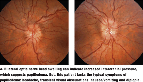
Upon dilation, however, the optic nerves appear elevated O.U. (figure 4). Bilateral optic nerve head (ONH) swelling can indicate increased intracranial pressure (ICP), which is suggestive of papilledema. But, this patient does not present with the typical symptoms of papilledema: headache, transient visual obscurations, nausea/vomiting and diplopia.
Diagnosis. First, check the patients blood pressure. Malignant hypertension, although rare in a healthy, young patient, can cause bilaterally swollen optic nerves.
 Second, if the blood pressure is normal, image the patient. Although this patient may have pseudotumor cerebriespecially if she is obese or on a medication that induces it (see Medications That Can Cause Pseudotumor)other causes of edematous optic nerves must be ruled out.5 As soon as possible, order the patient an MRI and an MRV (magnetic resonance venogram) of the brain. This combination is rapidly becoming the standard of care for evaluating bilaterally swollen optic nerves. The MRV is added to evaluate the venous system of the brain and to rule out a sinus thrombosis that is usually missed with an MRI. Without this information, the patient with a sinus thrombosis could be misdiagnosed with the more benign pseudotumor cerebri.6
All of Debras imaging was normal, so we referred her to a neurologist for a lumbar puncture (LP) to evaluate the opening pressure and confirm the diagnosis of pseudotumor cerebri. The patient with pseudotumor may be asymptomatic or may complain of headaches upon waking, as well as nausea and/or vomiting. Visual acuity is usually normal, but the patient may report transient visual obscurations or diplopia secondary to an induced sixth-nerve palsy.7 Visual fields often show enlarged blind spots.
Treatment/Management. Pseudotumor cerebri is most commonly treated with oral Diamox (acetazolamide, Lederle), although it is contraindicated in patients with severe kidney or liver disease and sulfa allergies. The side effects include tingling and numbness in the extremities, a metallic taste and aplastic anemia.8 The neurologist started Debra on Diamox 500mg b.i.d. and followed up with imaging, lumbar punctures and bloodwork. (The dose of acetazolamide is decreased and then discontinued over six months to one year, depending on the patients response.) The optometrist can comanage by following visual acuity, visual fields and the resolution of ONH edema every three to six months. If diplopia presents, prescribe Fresnel prisms until the decreasing intracranial pressure resolves the diplopia.
If the MRI shows a tumor or other cause for decreased cerebrospinal fluid flow, refer to a neurosurgeon. If the MRV shows a sinus thrombosis, refer the patient to a neurologist emergently for anti-thrombotic treatment with heparin and/or warfarin.9
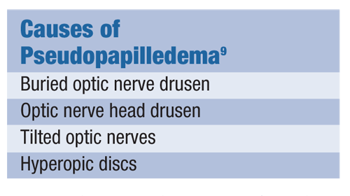 Discussion. When evaluating a patient with suspected papilledema, remember that other optic nerve conditions can mimic disc edema.10 (See Causes of Pseudopapilledema) Being able to differentiate between a truly swollen optic nerve and pseudopapilledema is often a challenge, even for the most experienced diagnostician. Blurred optic nerve head margins are not pathognomonic for optic nerve edema, so here are a few clinical questions that can help make the diagnosis:
Is the surrounding nerve fiber layer swollen?
Do the small optic nerve blood vessels become obscured at the edge of the disc?
Are there any disc hemorrhages?
Does prior documentation refute elevated optic nerve appearance?
Are the blind spots enlarged on visual field testing?
If the answer is yes to any of these questions, optic nerve edema is more likely.
Case 3: Bitemporal Hemianopia Indicates Pituitary Tumor
History/Diagnostic Data. Hannah is a healthy, 41-year-old black female who presents for reading glasses. An FDT visual field screening, done routinely on all patients, reveals a bitemporal hemianopia (figure 5). The patient is asymptomatic, and the remainder of the examination is normal.
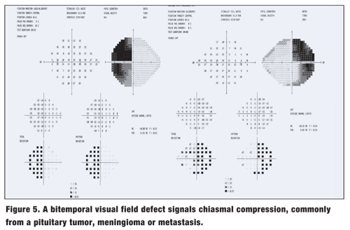
Diagnosis. Given this patients age and gender, she most likely has a pituitary tumor causing compression of the optic chiasm, resulting in a bitemporal defect. It is not unusual to be asymptomatic with a pituitary tumor, but some patients may report weight changes, menstrual irregularities or galactorrhea. Fortunately, most pituitary tumors are benign.
As with any patient who presents with a newly diagnosed neurological field defect, the first step is to order imaging. In general, non-glaucomatous neurological visual field defects are bilateral and respect the vertical midline in some fashion.11
Exceptions are unilateral and involve the optic nerve directly in conditions such as ischemic optic neuropathy, optic neuritis and compressive optic nerve lesions.12
In these cases, order an MRI of the brain and an MRI of the orbits. If an aneurysm is suspected, schedule an MRA (magnetic resonance angiography) to evaluate the arteries of the brain.
Treatment/Management. Hannahs MRI was positive for a large, benign pituitary tumor, and she was referred to a neurosurgeon. Following removal and/or treatment of the pituitary tumor, the surgeon will likely ask for follow-up visual fields to evaluate the effectiveness of the therapy. If a functional visual field defect remains permanent, the optometrist can consider the use of prisms to expand the visual field.
Discussion. With the increase in routine visual field screenings, optometrists are detecting defects earlier and more frequently. Lesions anterior to the chiasm produce a unilateral visual field defect. As in this patient, a bitemporal visual field defect signals chiasmal compression, commonly from a pituitary tumor, meningioma or metastasis. Postchiasmal conditions increase the congruity of the visual field.13 For example, ischemia or a tumor in the optic tract causes a contralateral homonymous hemianopia. A superior quadranopsia, or pie-in-the-sky defect, points to a contralateral temporal lobe problem, whereas an inferior quadranopsia (pie-on-the-floor) points to the contralateral parietal lobe.
Ischemia or bleeding from a stroke is the most common cause for a quadranopsia. Bilaterally enlarged blind spots signal swollen optic nerves from certain conditions, such as pseudotumor cerebri, and visual field defects with macular sparing warn us of occipital lobe involvement.14
How do you order an MRI? Simply write, MRI of brain with and without contrast or MRI of brain and orbits with and without contrast on a prescription pad with the patients name and your signature. (Most visual system pathway lesions are enhanced with an additional contrast study.) Include a tentative diagnosis and/or reason for imaging, such as bitemporal visual field defect or optic neuritis O.S.
Case 4: Binocular Diplopia And Cranial Nerve Palsy
History. James is a 57-year-old black male with a 15-year history of type 2 diabetes and hypertension. He presents complaining of a sudden onset of double vision for the past three days. The diplopia is horizontal and constant, and disappears when covering either eye. His last eye exam was four months prior, at which time he was diagnosed with non-proliferative diabetic retinopathy without clinically significant macular edema O.U.
Diagnostic Data. Preliminary testing was normal, except for a constant esotropia O.D. The retinal exam was unchanged from the previous visit.
Diagnosis. Based on the patients systemic conditions, his age and his constant diplopia, we suspected a microvascular-induced cranial nerve palsy. An MRI of the brain and orbits verified multiple areas of small-vessel ischemia in the brain.
Treatment/Management. The decision to order imaging in suspected ischemic nerve palsies is controversial, unless the diplopia persists for more than three months.15 Still, an MRI of the brain and orbits is not unreasonable to rule out other causes of acute-onset diplopia, such as compressive lesions, aneurysms, MS, thyroid disease, etc. If the MRI is normal or shows only small vessel disease, patients such as James can be reassured that the diplopia will likely resolve without treatment in three to six months. Unilateral patching or Fresnel prisms can be prescribed during this time.
Discussion. When evaluating diplopia, first determine if the double vision is binocular or monocular. Binocular diplopia signals an ocular muscle imbalance and disappears when one eye is covered.
Monocular diplopia, on the other hand, persists even when one eye is covered.16 Monocular diplopia originates from ocular conditions such as uncorrected refractive error, cataracts, dry eye, epiretinal membranes and corneal irregularities such as keratoconus.
Assuming the diplopia is binocular, the next important question is whether the diplopia is constant or intermediate. Constant diplopia is more often associated with cranial nerve palsies caused by vasculopathic conditions, pseudotumor and compressive lesions.17 Intermittent diplopia, on the other hand, is more common with a phoria breakdown, multiple sclerosis, myasthenia gravis and thyroid disease.17
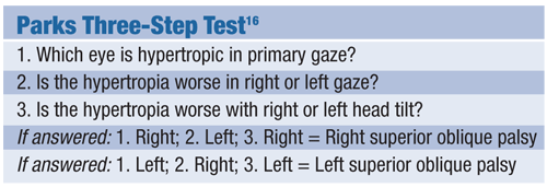 A third important question: Is the binocular diplopia vertical, horizontal or a combination? With microvascular muscle palsies, horizontal images and an esotropia point to a sixth-nerve palsy.15 The diplopia worsens when looking toward the affected side. Vertical images suggest a fourth-nerve palsy that can be supported by the Parks Three-Step Test (see Parks Three-Step Test).16 A third nerve palsy generally produces a combination of vertical and horizontal diplopia and often presents with additional signs, such as ptosis and a down-and-out appearance.18 Although head turns and tilts can help diagnose a congenital or long-standing nerve palsy, a patient with an acute, constant cranial nerve palsy will generally seek an evaluation before a head turn or tilt presents.
Neuro-optometric conditions can present with a variety of ocular signs and symptoms, ranging from optic disc edema and an APD to visual field defects and complaints of diplopia. Although there are certainly a myriad of neurological conditions with varying diagnoses and elaborate exceptions, the cases in this article are a few of the types most frequently encountered by optometrists.
These examples demonstrate the importance of examining the patient systematically, discussing any related systemic conditions, reviewing the signs and symptoms, and considering the patients demographic information, such as age, race and gender, in order to make the proper diagnosis.
Dr. Autry practices in a referral center in
1. Rosen ES, Thompsom HS, Cumming WJ, et al. Neuro-ophthalmology. 1st ed.
2. Karpecki PM. Studies provide new clues to AION. Rev Optom 2002;139(9):100-101.
3. Kanski JJ. Clinical Ophthalmology. 3rd ed.
4. Kunimoto DY, Kanitkar KD, Makar MS, ed. Wills Eye Manual. 4th ed.
5. Ang ER. Pseudotumor cerebri secondary to minocycline intake. J Am Board Fam Pract 2002;15(3):229-233.
6. Higgins JNP, Gillard JH, Owler BK, et al. MR venography in idiopathic intracranial hypertension: unappreciated and misunderstood. J Neurol Neurosurg Psychiatry 2004;75:621-625.
7. Friedman NJ, Pineda R,
8. Duramed Pharmaceuticals, Inc. Diamox package insert.
9. Allroggen H, Abbott RJ. Cerebral venous sinus thrombosis. Postgrad Med J 2000;76:12-15.
10. Karam EZ, Hedges TR. Optical coherence tomography in retinal nerve fiber layer in mild papilledema and pseudopapilledema. Br J Ophthalmal 2005 Mar;89(3):294-8.
11. Rosen ES, Thompsom HS, Cumming WJ, Eustace P, eds. Neuro-ophthalmology, 1st ed. London: Mosby, 1998:14.1-14.13.
12. Shikishima K, Kitahara K, Mizobuchi T, et al. Interpretation of visual field defects respecting the vertical meridian and not related to distinct chiasmal or postchiasmal lesions. J Clin Neuroscience 2006;13(9):923-928.
13. Purves D, et al. Neuroscience. Visual Field Pathway. 2nd ed.
14. Alexander LJ. Primary Care of the Posterior Segment. 2nd ed.
15. Shakya S, Agrawal JP, Ray P. Profile of isolated sixth cranial nerve palsy: A hospital based study. J Neuroscience 2004;1:32-35.
16. Danchaivijitr C, Kennard C. Diplopia and eye movement disorders. J Neurol Neurosurg Psychiatry 2004 Dec;75 Suppl 4:iv24-31.
17. Morris RJ. Double vision as a presenting problem in an ophthalmic causalty department. Eye 1991;5:124-9.
18. von Noorden GK. Binocular Vision and Ocular Motility. 4th ed.

