 Q: What higher-order aberrations are expected following DSEK and what are the remedies, if any?
A: First of all, it is important to note that Fuchs’ dystrophy patients have a higher amount of anterior corneal higher-order aberrations (HOAs) when compared to normal corneas. It is speculated that anterior stromal haze may play a role, and should not be ignored as a source of decreased visual acuity after DSEK.1
“The DSEK procedure seems to have little effect on anterior corneal HOAs, but does induce posterior HOAs, and hence total-eye HOAs,” says Richard Mangan, OD, Center Director at the Eye Center of Richmond, a multispecialty comanagement practice in Indiana and Ohio. A 2011 study found trefoil and quadrefoil were the predominant HOAs that emerged after DSEK.2
There was a correlation between increased HOAs and graft thickness 24 months after DSEK, and thicker grafts were also associated with more graft folds.2 The researchers also determined that there was little association between total HOAs and BCVA.
“What is more likely to contribute to decreased BCVA after DSEK is host-Descemet remnants at the donor-to-host interface, irregular graft thickness, interface reflection and/or donor contraction,” Dr. Mangan says. “In the event of poor BCVA after DSEK, the optical quality of the transplanted cornea can be improved by careful removal of the DSEK graft, and secondary application of a DMEK graft. Most consider this a better option than proceeding with a full-thickness transplant or PKP.”
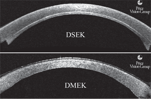
Cross-sectional images of DSEK vs. DMEK corneal grafts taken with AS-OCT. Photo: Francis W. Price, Jr., MD
In DMEK, a patient’s Descemet membrane and dysfunctional endothelium is replaced with the healthy endothelium and Descemet membrane from a donor cornea without the addition of any donor stromal tissue.
“One reason posterior surface HOAs increase after DSEK is because the procedure includes posterior donor stromal tissue, resulting in variable thickness,” says Francis W. Price, Jr., MD, founder and medical director of Price Vision Group in Indianapolis, Ind. “Since DMEK does not include any donor stroma, it is uniform and much thinner than a DSEK graft.”
Another cause of posterior surface HOAs is that no attempt is made to match the curvature of the donor and recipient corneas, so folds or wrinkles—either micro or macro—can develop as the graft conforms to the back of the host cornea.
“The visual impact is significantly greater with DSEK than DMEK because a DMEK graft is so much thinner,” Dr. Price says. “Thickness variations and folds or wrinkles in the graft result in substantially increased posterior corneal HOAs with DSEK.”
In contrast, the posterior corneal surface after DMEK more closely resembles that in age-matched control eyes, which is one reason DMEK provides superior visual results.3,4 In fact, in a series of patients who had DSEK in one eye and DMEK in the other, 83% preferred the vision in the DMEK eye.5
Many patients achieve excellent visual results with DSEK, but if they’re not satisfied with their vision after DSEK and the anterior surface is fine, then repeating the surgery with DMEK is the best treatment.6
1. Patel SC, Baratz KH, Maguire LJ, et al. Anterior corneal aberrations after Descemet’s stripping endothelial keratoplasty for Fuchs’ endothelial dystrophy. Opthalmology. 2012 Aug;119(8):1522-9.
2. Seery LS, Nau CB, McLaren JW, et al. Graft thickness, graft folds, and aberrations after descemet stripping endothelial keratoplasty for fuchs dystrophy. Am J Ophthalmol. 2011 Dec;152(6):910-6.
3. Rudolph M, Laaser K, Bachmann BO, et al. Corneal higher-order aberrations after Descemet’s membrane endothelial keratoplasty. Ophthalmology 2012 Mar;119(3):528-35.
4. Price MO, Giebel AW, Fairchild KM, Price FW Jr. Descemet’s membrane endothelial keratoplasty: prospective multicenter study of visual and refractive outcomes and endothelial survival. Ophthalmology. 2009 Dec;116(12):2361-8.
5. Guerra FP, Anshu A, Price MO, Price FW. Endothelial keratoplasty: fellow eyes comparison of Descemet stripping automated endothelial keratoplasty and Descemet membrane endothelial keratoplasty. Cornea. 2011 Dec;30(12):1382-6.
6. Ham L, Dapena I, van der Wees J, Melles GR. Secondary DMEK for poor visual outcome after DSEK: donor posterior stroma may limit visual acuity in endothelial keratoplasty. Cornea. 2010 Nov;29(11):1278-83.
