 |
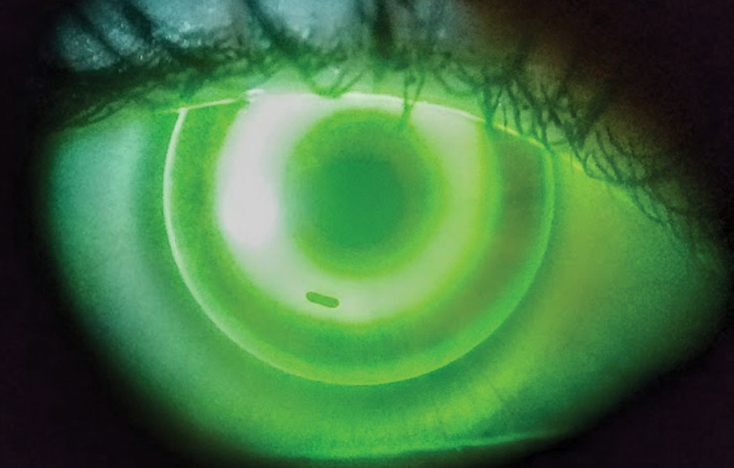
| Automatic OCT segmentation helps detect changes in the eyes of ortho-k patients. Photo: Dan Fuller, OD. Click image to enlarge. |
Many have used various methods to measure the geometrical changes that occur in the cornea due to orthokeratology (ortho-K) lens wear, including the thickness changes in the stroma and epithelium and the radius of curvature changes in the cornea's anterior and posterior surfaces. While these methods were largely successful in tracing the anterior and posterior surfaces, it remained difficult to detect the inner surfaces, and some methods involved manual segmentation steps. Researchers based in the University of Liverpool have developed and tested a new automatic segmentation method to trace the anterior corneal surface, epithelial posterior surface and posterior corneal surface in OCT scans.
A total of 45 right eyes from 52 participants between eight and 18 years old were monitored before and after one month of uninterrupted overnight ortho-K lens wear. Tomography was obtained using optical OCT and rotating Scheimpflug imaging (Oculus Pentacam). The researchers created a custom-built MatLab code for automatic segmentation of corneal OCT images and used it to assess changes in epithelial thickness, stromal thickness, corneal and stromal profiles and radii of curvature before and after one month of the evaluation period. The measurements were taken across the center of the cornea along both the horizontal and vertical meridians.
The study presented evidence that the corneal anterior surface experienced significant flattening in the central region after ortho-K lens wear. In the central area (0mm to 2mm diameter), the epithelium thinned by 12.8µm (23.8% on average) after one month of ortho-K lens wear. In the paracentral area (2mm to 5mm diameter), the epithelium thinned nasally and temporally by 2.4µm (4.5% on average). The researchers proposed that the thickness increases in the paracentral area could be due to the deformation of epithelial cells caused by the negative pressure exerted by the ortho-K lens in the reverse curve zone.
The stroma thickness increased in the central area by 4.8µm. The radius of curvature of the central corneal anterior surface increased by 0.24mm (3.1%) along the horizontal meridian and by 0.34mm (4.2%) along the vertical meridian. There were no significant changes in the anterior and posterior stromal radius of curvature.
The researchers concluded that they have established a method to automatically detect the three external and internal corneal surfaces in OCT scans. They believe that the surface detection results for 45 patients (180 OCT images) indicated the suitability of this method in segmenting corneal images.
“The method provides advantages in automation and validation in a relatively large patient population,” they noted. “To further enhance the new method, denoising of original OCT images and auto-detection of other corneal interfaces should be considered in future work.”
Ran Z, Moore J, Jiang F, et al. A new approach for quantifying epithelial and stromal thickness changes after orthokeratology contact lens wear. R Soc Open Sci. 2021;8(12):211108. |
