 |
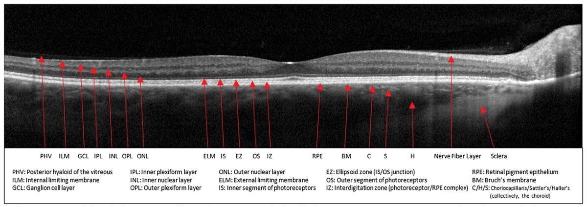
Geographic atrophy causes incorrect automated OCT segmentation in a high proportion of eyes with extensive outer retinal atrophy. Photo: Sara Weidmayer, OD. Click image to enlarge. |
Taking patients’ comorbidities into account is key when diagnosing and managing disease— especially so when glaucoma and age-related macular degeneration coexist. A recently published study reported that age-related macular degeneration can alter retinal topography and affect topography devices’ segmentation accuracy of the inner retinal layers.
The observational cross-sectional study included 125 eyes with non-exudative AMD. Patients’ drusen were automatically segmented on macular OCT B-scans using a publicly available, validated deep-learning approach. The inner plexiform layer-inner nuclear layer boundary was segmented with the OCT’s software.
The researchers reported drusen sizes ranging from 16µm to 272µm. Larger drusen were more likely to cause segmentation errors by compressing the inner macular layers. This may lead to “decreased thickness measurements that may be misinterpreted as atrophic changes of the inner retina,” they explained in their paper.
They observed that automated segmentation had a 22% chance of failure if drusen height was between 145µm and 185µm, with failure most likely with drusen heights greater than 185 µm. Additionally, they found that segmentation failed 36% of the time when the drusen-to-total retinal thickness ratio was 0.45 or greater, if drusen height were normalized by the total retinal thickness.
On OCT, images were more likely to show displacement of the inner retinal layers in the presence of drusen heights above 176 µm and a normalized drusen height ratio of 0.5 or higher. A total of 87% of images with outer retinal atrophy had incorrect segmentation.
“Segmentation of the inner retinal layers remains accurate in eyes demonstrating drusen with small to medium heights,” the researchers wrote. “Geographic atrophy causes incorrect automated segmentation in a high proportion of eyes with extensive outer retinal atrophy.
“Clinicians should be cognizant of the effects of outer retinal disease on the inner retinal layers and consider our findings when interpreting the results of automated macular segmentation in eyes with outer retinal diseases such as nonexudative age-related macular degeneration,” they concluded.
Chew L, Mohammadzadeh V, Mohammadi M, et al. Measurement of the inner macular layers for monitoring glaucoma: confounding effects of age-related macular degeneration. Ophthalmol Glaucoma. June 21, 2022 [Epub ahead of print]. |

