 |
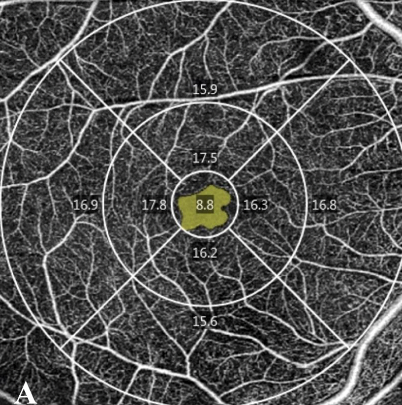
| The clear, detailed view of retinal microvasculature provided by OCT angiography offers a strong diagnostic adjunct without the invasive dye injection of traditionally used fluorescein angiography. This 6x6mm scan shows the vessel density of the different regions of the macula and the foveal avascular zone. Photo: Salongcay RP/Ophthalmic Research. Click image to enlarge. |
Routine retinal examinations are important for catching early signs of retinopathy in diabetic patients, and incorporating newer modalities can expedite detection and timely management. Adjunctive use of OCT angiography (OCT-A) to traditional modalities such as ultra-widefield color photos and ultra-widefield fluorescein angiography (FA) may improve the way clinicians detect and diagnose diabetic retinopathy and diabetic macular edema, according to a paper recently published in Ophthalmic Research that compared disease findings across these modalities.
In the prospective study, 114 eyes of 57 patients with diabetes were dilated and imaged with all three modalities. Ischemic areas seen on FA were identified with software that calculates the nonperfusion index (nonperfusion area divided by total area).
After exclusion for non-diabetic retinopathy findings or prior laser photocoagulation, 69 eyes were analyzed.
Here are the key findings:
- Diabetic retinopathy severity was associated with higher nonperfusion index, even after the researchers distinguished between rods and cones.
- Nonperfusion index correlated with diabetic macular edema and central subfield thickness in eyes with non-proliferative diabetic retinopathy.
- Fluorescein angiography macular nonperfusion correlated with all measures of nonperfusion index (total, rod, cone).
- Central vessel density and vessel perfusion correlated with presence of diabetic macular edema and central subfield thickness; and with macular nonperfusion in eyes with non-proliferative diabetic retinopathy.
- A larger foveal avascular zone correlated with decreased central vessel density and decreased central vessel perfusion.
Based on these findings, the researchers concluded that the three imaging modalities have complementary parameters. They wrote that the correlations among the findings each technology “provide clues on the pathogenesis and progression pattern of diabetic retinopathy and diabetic macular edema from which insights on clinical decision-making can be drawn.”
Salongcay RP, Aquino LAC, Salva CMG, et al. Comparison of diabetic retinopathy lesions identified using ultrawide field imaging and optical coherence tomography angiography. Ophthalmic Res 2023. [Epub June 28, 2023]. |
