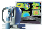
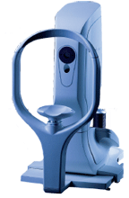
Spectralis,
 Heidelberg Engineerings Spectralis family includes five different models, OCT, OCTPlus, HRA, FA+OCT and HRA+OCT, which are specifically tailored to meet the particular demands of your practice.
All five models include TruTrack Image Alignment Technology, which enables the user to accurately document progression over time.
The HRA+OCT offers SD-OCT, FA, autofluorescence, red-free and infrared imaging modes, as well as single frame, multi-frame movie, stereo, mean, composite, real time and tomography imaging options. It takes 40,000 A-scans/second and features an axial resolution of 3.9m and a transverse resolution of 14m. Viewing options include color, spectrum, heat and high-frequency scales; grayscale; 2-D and 3-D modeling; thickness map; and thickness graph or progression.
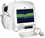
RTVue FD-OCT, Optovue
 The RTVue FD-OCT, by Optovue, offers posterior and anterior segment imaging, a retina and glaucoma normative database, progression analysis with vessel registration, RNFL and optic disc parameters, symmetry analysis, choroidal depth scanning and ganglion cell complex analysis.
"Spectral domain OCT with the Optovue for my glaucoma and ocular hypertension patients has been outstanding," says Robert Vandervort, O.D., of Omaha, Nebr. "From ganglion cell layer analysis to the NFL normative database to the anterior chamber documentation with the anterior segment module, there is just no comparison."
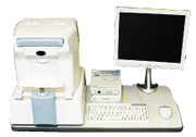
The RTVue scans OCT images at 26,000 A-scans per second, with 256 to 4,096 A-scans per frame. It has a depth resolution of 5.0µm in tissue and a transverse resolution of 15µm. Its scan range is 2.3mm deep and transverse, 2mm to 12mm. The fundus imager has a field of view of 32º by 23º, and requires a minimum pupil diameter of 3.0mm. The patient interface has a working distance of 22mm and a motorized focus range of -15.00D to +12.00D. The instrument has a footprint of 1,010mm by 520mm.
Cirrus HD-OCT, Carl Zeiss Meditec
 A new suite of OCT software applications for the Cirrus HD-OCT, by Carl Zeiss Meditec, has been approved by the U.S. Food and Drug Administration for commercial distribution.
The new suite of software includes three tools for glaucoma management:
-RNFL Normative Database, which helps the practitioner identify retinal nerve fiber layer loss.
-Anterior segment imaging, which visualizes the angle and measures of central corneal thickness.
-Guided Progression Analysis, which identifies statistically significant changes in RNFL thickness.
"Cirrus 4.0 takes OCT imaging to a whole new level," says Ike K. Ahmed, M.D., F.R.C.S.C., of the
The software package also includes new tools for retinal evaluation:
-Macular Change Analysis, which maps changes in macular thickness.
-Fovea Finder, which identifies the center of the fovea to help verify macular thickness measurements.
-Macular thickness normative data, which provides age-matched data for comparison.
The Cirrus OCT scan features an axial resolution of 5µm and a transverse resolution of 15µm. It takes 27,000 A-scans per second to a depth of 22.0mm at 1,024 points. Fundus imaging features a transverse resolution of 25µm and a 36º by 30º field of view. Focus can be adjusted from -20.00D to +20.00D, with both internal and external fixation. The technology can work with pupils as small as 2.0mm, though 3.0mm or larger is optimal. The instrument is 25.6” long by 17.3” wide by 20.9” high and weighs 83lbs.
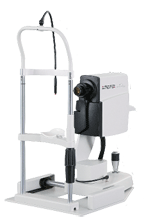
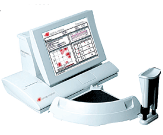
Park 1, Oculus
 The Park 1 diagnostic system, by Oculus, is a slim, ergonomic combination autorefractor, pachymeter and keratometer. It has a working distance of approximately 36mm and a 5 LCD color display for the practitioner. It is 20" high by 10.2" wide by 21.7" deep and weighs 26.4lbs.
The pachymeter function can measure from 200µm to 1,200µm and evaluates 600 data points in about one second. The keratometer function can measure from 9.00D to 99.00D and is accurate and reproducible to ±0.1D. The autorefractor function measures corneal vertex distances of 0mm, 12mm, 13.5mm and 15mm, sphere from -20.00D to +22.00D in increments of 0.12D at a corneal vertex distance of 12mm. Cylinder is measured in the same increment from 0.00D to 10.00D. Axis can be measured at each degree between 1º and 180º, and the minimum measurable pupil diameter is 2.5mm.
TRS-3100, Marco
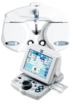 In partnership with Nidek, Marco introduces the TRS-3100, which follows in the footsteps of the TRS-5100 and features a new ergonomically-designed keypad and phoropter. The TRS-3100 is the first refraction system to offer a completely wireless interface with a Marco autorefractor and autolensmeter. The built-in IC Card Reader allows for this seamless integration and connection, without any cable logistics.
The responsive operator keypad makes refining sphere, cylinder and prism lenses smoother and faster than manual examinations. Also, during near point tests, power convergence and power prism diopter are adjusted automatically. Prism diopter can be adjusted independently for a more accurate and reliable measurement.
The TRS-3100 offers many of the same features as the TRS-5100. With the IC Card Reader and Marco Connect (Marcos proprietary product interface software, provided to all EMR companies), the practitioner can transfer data from pre-test equipment to the TRS-3100 and download the information to an EMR system.
Pascal Dynamic Contour Tonometer, Ziemer Group
 The Pascal Dynamic Contour Tonometer, by Ziemer, is a digital tonometer that takes a direct transcorneal measurement of IOP and detects ocular pulse amplitude. The tonometer uses the principle of contour matching, which negates any influence from central corneal thickness. It gathers 100 IOP values per second and records the dynamics of the values as a pulse wave, rather than one static figure.
The tonometer mounts onto a slit lamp. It is capable of measuring IOP between 5mm Hg and 80mm Hg with precision to 0.2mm Hg. It features an LCD display and requires SensorCap single-use tonometer covers. The sensor tip is concave and 7mm in diameter. The tonometer is 170mm high by 88mm wide by 40mm deep, and it weighs about 210g.
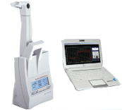
Ocular Response Analyzer, Reichert
 The Ocular Response Analyzer (ORA), by Reichert, utilizes a bi-directional applanation process to measure the biomechanical properties of the cornea and the IOP of the eye.
"Using a precisely metered, progressively escalating pulse of air, the ORA acquires corneal biomechanical data by quantifying the differential inward and outward corneal response to the air pulse over a time span of approximately 20 milliseconds," says Michael Sullivan-Mee, O.D., of Albuquerque, N.M.
The ORA takes 400 data samples to monitor the curvature of the cornea. It is also able to determine corneal-compensated intraocular pressurea measurement of IOP that is less affected by corneal properties than other methods of tonometry.
The ORA features a built-in ultrasound, pachymeter and a comprehensive patient management system.
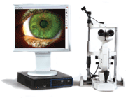
CellScreen Software and CellChek Specular Microscopes, Konan Medical
 Konans CellScreen software for the companys CellChek series of specular microscopes is designed to allow primary care doctors to determine the health of a non-medical patients corneal endothelium.
Early detection of corneal stress can minimize damage, as acute changes in cell morphology are mostly reversible. Awareness of endothelial abnormalities facilitates earlier care, which can minimize long-term effects.
"I was amazed at the number of patients in my practice who had corneal irregularities that were identified after I acquired my Konan specular microscope," says optometrist Mario Gutierrez of
Indications for screening include, but are not limited to:
-Contact lens wear.
-Long-term contact lens wear.
-Cataract surgery. -Refractive surgery.
-Glaucoma.
-Diabetes for more than 10 years.
-Prescription eye drops. -Dry eye.
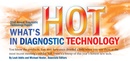
DigiVersal Slit Lamp System, Kowa Optimed
 The DigiVersal Slit Lamp Imaging System, by Kowa Optimed, adds image capture capability to slit lamps. The software package features live video display and video capture/replay. It can also capture stills from live or recorded video.
"Integrating the Kowa digital slit lamp and other digital equipment into my practice has allowed us to increase our efficiency and has helped us maintain better patient records," says David G. Kirschen, O.D., of the David Geffin School of Medicine at UCLA. "Once images are acquired with the digital slit lamp system, they are automatically placed into a patient folder so the images can be stored and presented to my patients as an educational tool."
The software uses a one-touch foot pedal system for live video image capture and includes a high-resolution Sony DFW-SX910 firewire digital camera, CD/DVD drive for non-magnetic digital backup, and toll-free telephone support.
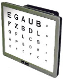
Smart System 20/20, M&S Technologies
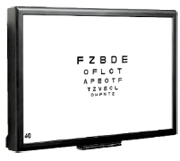 The Smart System 20/20, by M&S Technologies, is a computerized vision testing system that gives the practitioner the option to use polarized tests in addition to standard testsit comes with polarized glasses, shield overlay and three test options. The system includes myriad new vision tests, including E-ETDRS protocol, for clinical or trial-based use; sine wave grating contrast tests; dynamic astigmatic dials; ATS protocol, for clinical or trial-based use; and letter sizes to 20/1600.
The system features a 20" monitor, unlimited user-defined protocols, secondary calibration distance, fixation stimulus, desktop monitor display capabilities, Systemlink technology and full integration with Marco and Topcon autophoropters. The display may also be used for patient education purposes and is integrated with Eyemaginations software.
CVSi20 Computerized Vision System, Lombart
 The all-inclusive, Mac-based CVSi20 system by Lombart includes a 20" flat panel Apple LCD monitor and processor unit in one, wall-mounted piece. The 56-button remote allows you to select charts or chart combinations. Some of the charts included are Snellen letters, tumbling E, Landolt rings, numbers, HOTV, crowding bars, red/green testing, ETDRS and contrast testing as well as a variety of childrens charts. You can also add your own videos and images for patient education or fixation. Enhanced features include random character generation, single letter isolation, single line (vertical or horizontal) isolation, four-dot and fixation tests and included video clips for pediatric exam and fixation.
TRC-NW8, Topcon
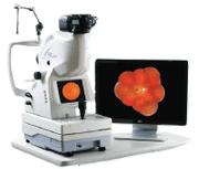 The TRC-NW8 non-mydriatic retinal camera, by Topcon, provides true red-free photography thanks to a built-in green filter. The TRC-NW8 features a Nikon digital SLR camera, lower flash intensity and fixation targeting, as well as alignment, auto-focus and auto-shoot aids.
The TRC-NW8 features 45 degrees
of coverage and a working distance of 40.7mm. It requires a pupil diameter of 4.0mm (3.3mm when using the small pupil diaphragm) for photography. It takes color and red-free photographs. The equipment can correct from -33.00D to +40.00D (with additional lenses).
The base has a side-to-side range of movement of 100mm, back-forth range of 46mm, and an up-down range of 30mm. The chinrest can move 67mm to accommodate patients. The TRC-NW8 weighs 24.5kg and is 272mm wide, 505mm long and 530mm high.
UltraVision Visual Acuity Tester, Woodlyn
 The UltraVision visual acuity tester and data access technology, by Woodlyn, allows practitioners to test acuity, record data and access all diagnostic data wirelessly from any location. UltraVision features HIPAA-compliant encryption that allows records to be securely transmitted.
"We've now introduced a "wow factor" to our patients when viewing the digital photos of their eyes," says Barry Basden, O.D., of
The UltraVision system allows the practitioner to retrieve any necessary data without having to leave the exam. It can be customized to fit any size practice or network, and is capable of reaching multiple locations.
The system also will streamline a practices ability to monitor patient records. "The diagnostic digital photos are much easier to track and bill as compared to hand drawings on file," says Benjamin Kachelman, O.D., of
Notal Vision & Sightpath Medical, Foresee PHP
 The Foresee PHP, by Notal Vision and distributed by Sightpath Medical, is approved for both the detection of choroidal neovascularization and the long-term monitoring of dry AMD.
"It is well known that early detection of choroidal neovascularization is our AMD patients best chance at preserving their sight," says Joseph J. Pizzimenti, O.D., of
The PHP performs a self-paced functional test of the 14-degree macular area based on hyperacuity--the patients sense of the relative spatial localization of multiple objects.
"Subtle functional changes in a patients central hyperacuity may indicate worsening of the condition and early formation of a choroidal neovascular membrane," Dr. Pizzimenti says. "The Foresee PHP's 82% sensitivity and 88% specificity give optometrists and their patients diagnosed with dry AMD the ability to track the condition more effectively."
Features of the Foresee PHP include Normative Database Analysis to aid in the early detection of CNV and quantitative automated threshold visual field test for the monitoring of AMD. The noninvasive exam takes from three to five minutes per eye. The user interfaced is intuitive and automated, and the reports provided to the doctor are clear and objective. They highlight chronological progression.
"One of the Foresee PHP's many benefits is its ease of use for both patients and technicians," says Dr. Pizzimenti. "The vast majority of my patients with early and intermediate AMD are able to perform the test with reliable results in about four minutes per eye. For those colleagues who see more than 10 patients per week with multiple drusen, I highly recommend the Foresee PHP."
