|
Often, gradual anatomical changes to the eyelids go unnoticed, or unremarked during exams, yet can significantly affect our patients' vision—and their self-perception. Blepharoplasty can be performed for cosmetic or medical reasons, both of which should be considered when talking to patients. The most common diagnosis leading to blepharoplasty is dermatochalasis, an age-related, progressive and bilateral eyelid droop resulting from redundant, loose eyelid skin and herniation of the orbital fat.
Who Benefits?
Dermatochalasis is often regarded as an independent condition; however, one study found that 51% of patients also complained of dry eye. Of those patients, 86% reported improvements in dry eye symptoms following upper blepharoplasty.1
Patients with significant dermatochalasis may lose a portion of their superior vision. Glaucoma patients with the condition may do poorly on visual field testing and present with a pseudo-altitudinal defect, which improves after the procedure.2
Many dermatochalasis patients who present to us without a measurable visual defect may be thinking about aesthetic concerns; so, be sure to consider this before writing off the idea that they may be interested in elective procedures.
Facial rejuvenation via blepharoplasty can also improve the aesthetics of both the brow and cheek. Don't be afraid to address these topics with patients—they will be glad you did.
Screening
Many patients who can benefit from a blepharoplasty may not know it, or assume the procedure is elective so they don't consider it. For example, a patient who has difficulty opening their eyes at the slit lamp is an optimal patient with whom to initiate a discussion.Obtain a thorough history regarding current systemic conditions and medications, and pay attention to a history of keloid scarring, and drugs or supplements that are anticoagulants. Screen patients for dry eye, glaucoma, pre-existing ptosis, lagophthalmos and thyroid eye disease.
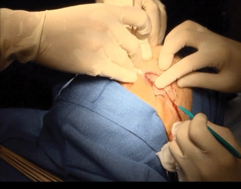 |
| During upper lid blepharoplasty, a crescent of skin and the underlying orbicularis muscle is excised to remove or trim prolapsing, preaponeurotic fat. |
The upper margin-reflex distance (MRD-1) indicates the dermatochalasis is severe if the distance from center of the pupil to the eyelid margin is 2.5mm or less. The superior, 36-point screening test with and without taping is standard to show the visual significance of dermatochalasis and is often used by insurers to determine coverage.
Surgical Overview
Blepharoplasty can be performed in-office under local anesthesia or under sedation at a surgical center. Upper lid blepharoplasty uses an external incision to create and remove a small crescent of skin, along with any prolapsing medial or central fat pads. Resection with absorbable sutures of retro-orbicularis oculi fat helps decrease the upper lid and lateral brow weight.
Lower lid blepharoplasty uses either an external incision below the lash line for dermatochalasis excision or an internal incision through the conjunctiva below the lid margin to conservatively remove fat.
Significant swelling and bruising is likely after surgery. Advise patients to use ice packs for 15 minutes each hour in the two to three days following surgery to reduce edema. Topical antibiotic ointment is applied twice daily until sutures are removed, and pain is managed with acetaminophen 1000mg. Vitamin E ointment can be massaged into the skin after suture removal to minimize scar formation. At the one-month follow up, assess positioning and symmetry between the eyelids.
  |
|
1. Vold S, Carroll RP, Nelson JD. Dermatochalasis and dry eye. Amer J Ophthalmol. 1993;115(2):216-20. 2. Alan KS, Wishart PK, Birch MK. Apparent glaucomatous visual field defects caused by dermatochalasis. Eye. 1997;11(Pt 5):682-6. |
