 A 77-year-old black female presented to the office as a new patient in July 2009. The patient’s daughter, an established glaucoma patient, accompanied her to the visit. The patient recently moved in with her daughter after moving from New York because of increased difficulty with independent living due to progressive visual impairment.
The patient was last seen in New York in April 2009, and had previously undergone cataract surgery in her right eye in July 2008. She reported minimal improvement in her vision following the surgery. Her ocular history was significant for bilateral trabeculectomy. The patient reported that she was diagnosed with glaucoma nearly three years earlier, but had “very bad vision for a while” in both eyes prior to the diagnosis.
At the time of her visit to my office, she was medicated with Lipitor (atorvastatin, Pfizer), furosemide, Aggrenox (aspirin/extended release dipyridamole, Boehringer-Ingelheim), dietary potassium, Diamox Sequels (acetazolamide, Lederle Laboratories) b.i.d., 1 drop Xalatan (latanoprost, Pfizer) h.s. O.D., 1 drop dorzolamide q.d. O.D. and 1 drop Combigan (brimonidine tartrate and timolol maleate, Allergan) b.i.d. O.D. She also recently received a third course of methotrexate.
Diagnostic Data
Her best-corrected visual acuity was 20/200 O.D. and 20/400 O.S., with no improvement upon pinhole or refraction. Pupils were reactive to light, with no afferent defect. The left pupil was irregular in shape and showed minimal movement on light stimulation.
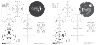
Our patient’s visual fields were significantly aberrant in both eyes (O.D. left, O.S. right).
Pachymetry readings were 542µm O.D. and 549µm O.S. Intraocular pressure measured 19mm Hg O.D. and 11mm Hg O.S. at 10:25 a.m.
Her corneas were clear in both eyes, with a clear corneal incision located temporally in her right eye. Both anterior chambers were deep and quiet. The left iris showed 270º of posterior synechia, with irregular pupillary ruff.
There was a surgical peripheral iridectomy located at 12 o’clock in the right iris, and two laser peripheral iridectomies in the left iris. Filtering blebs were located superiorly in both eyes. The bleb in the right eye was flat and avascular, but the bleb in the left eye was still well-formed and demonstrated no leak or blebitis.
Threshold visual fields were significantly aberrant in both eyes. The right field was constricted to the central 5º to 8º degrees, with a small temporal island of vision near the physiologic blind spot. The left field showed three 5º islands of vision approximately 25º to 30º from fixation, as well as a small central island below fixation. Threshold field testing times were in excess of 13 minutes in the right eye and 17 minutes in the left eye. Needless to say, the patient was fatigued following the field testing.
Upon dilated exam, the patient was pseudophakic in the right eye, with a well-centered posterior chamber IOL in the capsular bag and an opened posterior capsule. The crystalline lens in the left eye was characterized by 2+ nuclear sclerosis and cortical spokes in the visual axis. Bilateral posterior vitreous detachments were present.
Stereoscopic examination of the optic nerves demonstrated cup-to-disc ratios of 0.95 x 0.95 O.D. and 0.90 x 0.95 O.S. Our patient had complete loss of the neuroretinal rim in her right eye from 2 to 10 o’clock, as well as complete neuroretinal rim loss from 12 to 7 o’clock in her left eye. Both maculae were characterized by dry age-related retinal pigment epithelium granulation and disruption.
There was a significant epiretinal membrane (ERM) involving the foveal avascular zone in her right eye as well as a more subtle ERM located centrally in her left eye. Her peripheral retinal examination was remarkable for 360º of pigmented cystoid and reticular degeneration in both eyes.
Discussion
So, what options––if any––do we have to at least maintain this patient’s remaining vision? Can we even begin to speculate that her vision could be improved?
Her visual fields were affected significantly by her advanced glaucoma. Additionally, she had a dense cataract in her left eye and a failed filtering bleb in her right eye. Though the visual acuity was worse in her left eye, the neuroretinal rim was marginally healthier than that of the right.
An argument can be made that removal of the cataract could improve her vision either to or beyond the level of the right eye. However, in addition to her frail rim picture, I had two primary concerns about recommending cataract surgery: the presence of posterior synechiae and the patient’s use of methotrexate.
The existence of posterior synechiae in her left eye is a strong indicator of significant anterior chamber inflammation, which could be worsened by cataract removal. Additionally, the use of methotrexate, an anti-metabolite, suggests that the patient likely has recalcitrant rheumatoid arthritis. Given the patient’s history of auto-immune disease, any type of intraocular surgery could trigger an exaggerated postoperative inflammatory reaction.
Because of these risks for severe inflammation, the patient could require a prolonged postoperative period of steroids, which would then result in problems with IOP control. So, for these reasons, I elected not to proceed with cataract surgery.
One could also argue that the patient’s IOP in her right eye is not adequately controlled and that a second filtering procedure should be attempted. But, at her initial visit, it was impossible to tell if her visual field and neuroretinal rim loss had stabilized. Only time would tell if it has. For this reason, I continued her current therapy and asked her to return in one month for gonioscopy and Heidelberg Retina Tomograph-3 (HRT-3, Heidelberg Engineering) imaging.
As requested, the patient returned one month later. Her IOP was 18mm Hg O.D. and 12mm Hg O.S. Gonioscopy revealed 4+ open angles O.D. with a superior sclerostomy, and 3+ open angles O.S. with a superior sclerostomy. HRT-3 nerve scans supported the earlier diagnosis of severe bilateral neuroretinal rim thinning. She reported compliance with her medications.
Unfortunately, this patient’s long-term prognosis is rather bleak. As previously noted, surgeries to address the cataract in the left eye or the failed filter in the right eye pose significant risks to her ocular health. But, because she is tolerating the Diamox quite well, it is reasonable to believe that continued therapy may help sufficiently maintain her vision over the long term.
Still, periodic lab reports will need to be ordered to monitor electrolyte balances. Additionally, a close relationship with the patient’s internist must be maintained, because the concurrent use of an oral diuretic can negatively interact with the Diamox.
Should her IOP rise or her neuroretinal rim be further compromised down the road, the patient would be best served with either argon laser trabeculoplasty (ALT) or selective laser trabeculoplasty (SLT). In this patient’s case, ALT or SLT would likely decrease her IOP by a few points and have the least likelihood of side effects.
A retrospective analysis published in the American Journal of Ophthalmology in 2002 indicated that the presence of field loss at the time of initial diagnosis is one of the most important considerations in determining which glaucoma patients will proceed to blindness.1 Accordingly, our patient reported a rather recent diagnosis of glaucoma with several preceding years of poor vision. Additionally, a review of her medical records demonstrated significant field compromise at the time of initial diagnosis. And, unfortunately, our patient’s progression to visual impairment can continue, despite undergoing bilateral trabeculectomy.2 Finally, variability in IOP control is always a risk for progression to visual impairment.1,3,4
As with all glaucoma patients, you must meticulously evaluate the neuroretinal rims, visual fields and intraocular pressure at each visit. Should you observe deterioration, you must take the next management step. But eventually, in some patients, management options do, in fact, cease to exist––despite heroic therapy early in the process.
To date, our patient has not demonstrated additional progression. We’ll see how long that remains the case.
1. Oliver JE, Hattenhauer MG, Herman D, et al. Blindness and glaucoma: a comparison of patients progressing to blindness from glaucoma with patients maintaining vision. Am J Ophthalmol. 2002 Jun;133(6):764-72.
2. Ehrnrooth P, Puska P, Lehto I, Laatikainen L. Progression of visual field defects and visual loss in trabeculectomized eyes. Graefes Arch Clin Exp Ophthalmol. 2005 Aug;243(8):741-7.
3. Caprioli J, Coleman AL. Intraocular pressure fluctuation a risk factor for visual field progression at low intraocular pressures in the advanced glaucoma intervention study. Ophthalmology. 2008 Jul;115(7):1123-1129.e3.
4. Nouri-Mahdavi K, Hoffman D, Coleman AL, et al. Predictive factors for glaucomatous visual field progression in the Advanced Glaucoma Intervention Study. Ophthalmology. 2004 Sep;111(9):1627-35.
