Headaches frequently localize in and around the eyes, and patients who experience them on a recurring basis often present to you, their primary eye care provider, for help. There are myriad reasons why a patient is in your office discussing their headache, and arriving at the proper diagnosis isn’t as straightforward as you might think.
For example, although headaches are commonly associated with eyestrain, or asthenopia, these patients often describe vague feelings of visual discomfort rather than true head pain. Certain ocular pathologies result in chronic recurrent head pain as well, but these diagnoses are usually self-evident by gross observation, or are revealed through biomicroscopy or ophthalmoscopy. While knowing which ocular pathologies present with symptoms of head pain is important, headaches of ocular origin do not represent the vast majority of chronic recurring headaches.
Recurring headaches are broadly classified as primary or secondary. Secondary headaches are caused by an underlying pathology or structural abnormality and may be indicative of a potentially serious systemic or neurologic entity (Table 1).2-4 Fortunately, most patients have primary headaches, which are benign and are not caused by an underlying disease or structural problem.1,2 Although they may cause significant pain and disability, they are not dangerous. The two most common primary recurring headaches are tension-type headache and migraine.2
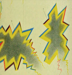 |
| This drawing by Joseph Babinksi vividly portrays the visual aura seen by individuals who suffer from migraine. Photo: Welcome Library. |
Because migraines are so common, it is important that you be ready to diagnose and manage patients who present with them. This review will help you better understand the etiology and pathophysiology of migraine and will walk you through clinical evaluation and management approaches that are integral to helping this patient population.
Signs and Symptoms
Researchers estimate approximately 10% of the population suffers from some form of migraine disorder.5,6 Most patients note that their migraines began in the early teens or, less commonly, in childhood.2 The onset of migraines is not common after age 40 and is rare after the age of 65.6 In addition, migraines have a strong family tendency, are more common in females, are influenced by hormonal factors and may be induced by certain triggers (Table 2).7,8
Diagnosis
Migraine is a diagnosis of exclusion; all other potential pathologies related to secondary headache must be eliminated through examination before diagnosing migraine. Patients with migraines usually present with a stereotypical symptom complex. The head pain is:
- Described as throbbing, pulsating and pounding.
- Usually unilateral upon onset but may spread to the other side of the head.
Accompanied by a variety of physical symptoms that often include lack of appetite, nausea, vomiting, diarrhea, cramping, photophobia, hyperacusis leading to sonophobia, excessive sweating (diaphoresis), vertigo and extreme fatigue.7
Patients can be quite sick during these episodes and often refer to them as “sick headaches.” Following the intense head pain, there may be sustained muscle contractions in the neck and shoulders.
The intense pounding headache, with the attendant constitutional signs and symptoms, usually lasts from three to seven hours, but it may persist for up to 72 hours. A migraine without aura lasting more than 72 hours is known as status migrainosus. Chronic migraine fulfills the diagnostic criteria for typical migraine, but occurs for 15 days or more per month for more than three months.7,8
Table 1. Secondary Headaches Requiring Additional Investigation | |
| Secondary Headache | Possible Etiology |
| Recurrent headaches in patients younger than age five. | Arteriovenous (AV) malformation. |
| Recurrent headaches in patients older than 50. | Cranial arteritis, mass lesion. |
| Abrupt-onset, acutely painful headache (“worst headache of my life”). | Subarachnoid hemorrhage. |
| Headaches of recent origin that are becoming increasingly more painful. | Mass lesion; subdural hematoma. |
| Headaches with concomitant fever, stiff neck, vomiting, cutaneous rash. | Meningitis, encephalitis, Lyme disease, collagen vascular disease. |
| Headaches associated with non-remitting neurological signs or symptoms such as papilledema, vertigo, seizures, personality changes. | Mass lesion, AV malformation, increased intracranial pressure, encephalitis, meningitis. |
| Headaches abruptly after bending, coughing, exertion or Valsalva. | Mass lesion, subarachnoid hemorrhage. |
| Headaches abruptly after head trauma. | Epidural or subdural hematoma. |
| Headaches associated with systemic cancer or HIV. | Metastasis, opportunistic neurologic infection. |
| Headaches during pregnancy or postpartum. | Venous sinus thrombosis. |
Pathophysiology
Although the underlying mechanisms of migraine are not completely understood, researchers theorize that, subsequent to stress or nonspecific stimuli, platelets aggregate and release the neurotransmitter serotonin. In turn, the neurotransmitter causes vasoconstriction of certain blood vessels that supply the base of the brain and other posterior brain vasculature. The regional reduction in blood flow causes a cortical spreading depression (CSD) that proceeds anteriorly. The CSD results in the prodromal signs and symptoms, as well as the aura. Additionally, the reduced blood supply results in local tissue abnormalities such as hypoxia, acidosis and carbon dioxide buildup. The parenchymal arteries dilate in response to increased local tissue demands, activating pain-modulating afferents within the trigeminal nerve and trigeminal vascular system at the base of the brain.
Activation of the trigeminal vascular system by CSD stimulates neurons in dural blood vessels to release plasma proteins and pain-generating substances such as calcitonin gene-related peptide (CGRP), substance P, vasoactive intestinal peptide and neurokinin A. These sterile inflammatory substances produce edema, sensitize cranial pain receptors and lower the pain threshold, leading to the characteristic pounding headache.9-12
Before, During and After
About 30% to 40% of migraine patients report a prodromal phase that occurs from 24 to 48 hours prior to the onset of the migraine.6 This should not be confused with the migraine aura, which is more proximal to the headache. Prodromal signs and symptoms may include aphasia, mood changes, diarrhea, excessive urination fatigue, repetitive yawning, food cravings, increased thirst and problems sleeping and concentrating.
Additionally, many migraine patients experience a postdromal phase, sometimes called the migraine hangover, which can last several hours and includes malaise, fatigue, poor concentration and altered mood.
Several variations of migraine exist, each with their own characteristic symptoms:
Migraine with aura (classic migraine). Approximately 10% to 15% of migraine sufferers experience auras that precede the onset of the headache.13 The visual phenomena develop gradually and last less than 60 minutes. They appear as flickering, flashing or scintillating positive scotomas that may surround an area of reduced vision. They may also appear as sparkles or heat waves that have a zigzag appearance. The scintillations often begin near the center of the visual field and expand slowly as they move outward toward the periphery. The light flashes are usually hemianopic and manifest contralateral to the headache. Occasionally, patients experience visual hallucinations such as macropsia or micropsia.
Some patients experience gradually developing, non-visual, sensory and motor auras, including numbness or tingling in the arms and hands, paresthesias of the tongue resulting in slurred speech and, rarely, vertigo and ataxia. Visual and non-visual auras precede the headache by 10 to 20 minutes. It is important to emphasize that both visual and non-visual auras build up gradually and are reversible in most cases. In contradistinction, the neurologic signs and symptoms of stroke occur suddenly and are usually non-reversible.
Migraine without aura (common migraine). Migraine without aura affects approximately 85% of migraine sufferers and is characterized primarily by headache and attendant gastrointestinal signs and symptoms.13 Patients do not manifest focal, gradually-developing symptoms of aura.
Table 2. Characteristics of the Migraine Patient7,8 |
| Childhood or early teens |
| Strong family history |
| More common in females |
| Increase in frequency during menstruation |
| Increase in frequency with birth control pills |
| May decrease in frequency during pregnancy |
| Usually decrease in frequency after menopause |
| May be triggered by foods containing tyramine, nitrites or monosodium glutamate |
| May be triggered by mental stress, exertion, dazzling light or high altitude |
Retinal migraine. Also called ophthalmic or ocular migraine, this is a fairly common cause of transient monocular blindness in young adults.14,15 This disorder is manifested by recurrent attacks of unilateral visual disturbance or blindness lasting from minutes to one hour. The visual phenomena may be associated with the typical migraine headache or with minimal or no headache (acephalgia). Patients often describe a developing central scotoma that gradually enlarges to produce total unilateral visual loss. Postural changes, exercise and the use of oral contraceptive agents may precipitate attacks.15 The visual symptoms are totally reversible.8,15
Retinal migraine is thought to result from transient vasospasm of the choroidal or retinal arteries. Rarely, when patients with retinal migraine are examined during an attack with visual loss, optic pallor or narrowing of the retinal vessels can be seen.14 A history of recurrent attacks of transient monocular visual disturbance or blindness, with or without a headache and without other neurologic symptoms, is suggestive of retinal migraine. A personal or family history of migraine confirms the diagnosis along with exclusionary examination and testing. Retinal migraine must be differentiated from ocular or vascular causes of transient monocular blindness such as carotid artery disease and coagulation disorders.
Aura without headache (acephalgic migraine). During an acephalgic migraine, the patient experiences visual, sensory and motor auras without headache. This phenomenon is characterized by repeated episodes of visual, sensory and motor neurologic symptoms that develop gradually, last approximately one hour or less, are completely reversible. This presentation can occur in patients of any age but often occurs in older patients who have had a history of migraine with aura at an earlier age.8 Aura without headache must be differentiated from transient ischemic attacks, occipital lobe seizures and temporal lobe seizures.
Migraine variants. This term is not used in the classification of the International Headache Society, but includes those forms of migraine that are not typical of migraine with or without aura.8 Migraine variants often have significant neurologic manifestations associated with the headache. The neurologic signs and symptoms may precede or occur coincident with the headache and may persist for hours or days after the headache subsides. The headache is often not the most important feature of these rare migraines, and it is usually shorter in duration and less severe than in the typical migraine.
• Hemiplegic migraine is an especially rare form of familial migraine that often starts in childhood and disappears in adulthood. These recurrent headaches are associated with unilateral hemiparesis or hemiplegia. Although the neurologic deficit usually resolves before the headache, occasionally the problems persist for days to weeks. Most people with hemiplegic migraine have inherited a dominant gene mutation from a parent who also suffered from the condition.16-18
• Basilar-type migraine, formerly known as Bickerstaff’s syndrome, is, in essence, a migraine with aura, with the symptoms originating from the brainstem and affecting both hemispheres of the brain at the same time. Patients present with fully reversible symptoms of vertebral basilar vascular insufficiency, which usually precede the headache by an hour. However, there is no motor weakness. The most common symptoms are vertigo and dizziness, but symptoms may also include diplopia, hemianopsias, ataxia, tinnitus, decreased hearing, nausea, bilateral paresthesias, syncope and loss of consciousness.19
• Ophthalmoplegic migraine is a rare disorder that typically starts in childhood. It is characterized by repeated typical migraines associated with paresis of one or more extraocular muscles. Most commonly the oculomotor nerve is affected, but the abducens or trochlear nerves may also be involved. Brain scans do not reveal any intracranial masses; however, several studies show reversible thickening or contrast enhancement of the cisternal portion of the oculomotor nerve on MRI.20-22 This finding suggests that the ocular muscle palsies caused by oculomotor nerve involvement may be due to recurrent inflammation.
Other rare primary migraines are listed in Table 3.
Table 3. Rare Primary Headaches2,7 | |
| Headache | Clinical Characteristics |
| Cluster headache | Sudden recurrent headache localized around one eye; headaches occur in clusters of one to five per day for a period of four to six weeks; each headache lasts from 20 to 90 minutes and is accompanied by ipsilateral facial sweating, lacrimation, nasal and conjunctival congestion, miosis and ptosis. |
| Hemicrania continua | Continuous unilateral headache of moderate intensity lasting up to three months; occasional exacerbations of severe pain accompanied by ipsilateral nasal and conjunctival congestion, ptosis and miosis. |
| Primary stabbing headache (“ice pick headache”) | Recurrent episodes of stabbing pain in or around one eye, lasting several seconds. |
| Primary cough headache | Sudden headache that lasts several minutes after coughing, sneezing or straining. |
| Primary exertional headache | Throbbing, pulsatile pain starting during or after exercise. |
| Primary sex headache | Dull, bilateral headache that starts during sexual activity and becomes worse during orgasm. |
| Hypnic headache | Short-lasting headache that starts a few hours after falling asleep; may recur several times during the night; often awakens the patient from sleep. |
Treatment
Intervention should have a three-pronged approach: general measures with analgesia, abortive measures to prevent an acute attack and prophylactic therapy to prevent recurrence.
General measures include minimizing or avoiding the conditions and agents that trigger migraine attacks. In particular, the patient should attempt to avoid dietary items with vasoactive properties. Refraining from using birth control pills and minimizing mental stress and physical fatigue has also been found to be helpful. Clinicians should also investigate possible sleep dysregulation, such as obstructive sleep apnea, periodic limb movement disorder, insomnia and hypersomnia, since studies suggest migraines may be consequent to, or aggravated by, these disorders.23-25
Abortive measures are pharmacological and include both simple analgesics, such as acetaminophen, and nonsteroidal anti-inflammatory drugs (NSAIDs). Simple analgesics, such as acetaminophen, can help by raising the pain threshold. Nonsteroidals such as aspirin, naproxyn, ibuprofen and indomethacin inhibit prostaglandin synthesis and reduce pain induced by the trigeminal-vascular system.26
The mainstay abortive pharmaceutical treatment for more severe migraines historically has been ergotamine tartrate, which is a derivative of ergot alkaloids and is a potent vasoconstrictor. It specifically counteracts the dilation of various branches of the trigeminal-vascular system affected in migraine. To be effective, ergotamine tartrate must be administered during the painless, pre-headache phase or soon after onset.
Triptans, first introduced in 1992, are currently considered the first-line treatment for moderate to severe migraines.28 These serotonin receptor agonists inactivate receptors located on the peripheral trigeminal nerve terminals that supply pain-sensitive vascular meningeal structures. Additionally, these drugs block the neuropeptide-mediated inflammatory response after trigeminal stimulation and may also block transmission in trigeminal neurons.30,31
Triptans are contraindicated if the patient is also taking selective serotonin reuptake inhibitors (SSRIs). These drugs cause serotonin to remain in an elevated concentration, and the addition of triptan could result in a serotonin syndrome consisting of anxiety, flushing, paleness, tremors, increased heart rate, fever, diarrhea and vomiting. Additionally, vasoconstrictive agents such as triptans and ergotamine tartrate should be avoided for retinal and basilar-type migraines.32
Prophylactic treatment is warranted when migraine attacks are frequent and the patient’s lifestyle is disrupted. Among the most effective prophylactic drugs are the beta-adrenergic blockers (e.g., propranolol, atenolol, metoprolol) and the calcium channel blockers (e.g., verapamil, diltiazem, amlodipine, nefedipine).33-35 The beta-adrenergic blockers inhibit platelet aggregation and reduce the liberation of prostaglandins and other sterile inflammatory substances that induce pain. The calcium channel blockers are thought to prevent intracranial vasoconstriction and the spreading of migrainous cortical depression.
Other useful prophylactic drugs include tricyclic antidepressants (TCAs) such as amitriptyline and certain antiepileptic drugs such as Depakote (valproic acid, Abbott Pharmaceuticals), Topamax (topiramate, Janssen Pharmaceuticals) and Neurontin (gabapentin, Pfizer). TCAs act primarily as serotonin-norepinephrine reuptake inhibitors. The resultant elevated concentration of serotonin and norepinephrine inhibit pain-inducing nerve impulses from the trigeminal-vascular system.35 Depakote, Topamax and Neurontin are believed to enhance gamma-aminobutryic acid neurotransmission, which may suppress events related to migraine.36-39
Injections of Botox (onabotulinum toxin A, Allergan) into the glabellar, frontalis and temporalis muscles have proven to be an effective prophylactic for migraine patients who do not respond to other therapies.40,41 Botox directly decreases the release of pain mediators, including substance P and CGRP from trigeminal sensory afferent terminals.42 Additionally, Botox inhibits the release of glutamate, which helps stimulate the release of pain mediators. Finally, Botox inhibits sensitization of central trigeminal vascular neurons. Research shows central sensitization is an integral factor in the development and progression of migraines.40-42
The FDA approved the transcutaneous electrical nerve stimulation (TENS) device, the first prophylactic medical device for migraines in adults, in 2014. It fits across the forehead and over the ears and stimulates the trigeminal nerve with a self-adhesive electrode in the center of the forehead. In one study, the device reduced the number of migraine days per month and reduced the amount of abortive medication use.43
Conclusion
Properly diagnosing your patients with typical or atypical migraine syndrome calls for a systematic approach. A detailed and relevant history is the most important factor, and careful questioning often reveals a particular headache profile that allows you to make the diagnosis. Ask about onset, time of day, location, frequency, duration, quality and severity, prodromes, precipitating factors, associated symptoms, family history, medical history and response to therapy. A thorough ocular health examination will help you rule out anterior segment, retinal or neuro-ophthalmologic pathologies.
You should be particularly concerned if the history, examination or both suggest the headaches are secondary to an underlying pathology or structural abnormality, in which case an appropriate referral is mandatory.
Dr. Blaustein is associate professor at Pennsylvania College of Optometry and Chief of Optometry Services at the Veterans Affairs Medical Center in Coatesville, PA.
|
1. Latinovic R, Guillifird M, Ridsdale L. Headache and migaine in primary care: consultation, prescripition, and referral rates in a large population. J Neurosurg Psychiatry. 2006;3(1):385-7. 2. Larner AJ. Guidelines for primary headache disorders in primary care: an “intervention” study. Headache Care. 2006;3(1):1-2. 3. Lamont AC, Alias NA, Win MN. Red flags in patients presenting with headache: clinical indications for neuroimaging. Br J Radiol. 2003 Aug;76(908):532-5. 4. Goadsby PJ, Raskin NH. Chapter 14. Headache. In: Longo DL, Fauci AS, Kasper DL, Hauser SL, Jameson J, Loscalzo J. eds. Harrison’s Principles of Internal Medicine, 18e. New York, NY: McGraw-Hill; 2012. 5. Stovner L, Hagen K, Jensen R, et al. The global burden of headache: a documentation of headache prevalence and disability worldwide. Cephalagia 2007;27(3):193-210. 6. Tepper SJ, Dahlof CG, Dowson A, et al. Prevalence and diagnosis of migraine in patients consulting their physician with a complaint of headache: data for the Landmark Study. Headache. 2004;44(9):856-64. 7. Olesen J. The international classification of headache disorders. Headache. 2008;48:691-3. 8. Headache Classification Committee of the International Headache Society. The international classification of headache disorders, 3rd edition (beta version). Cephalagia 2013;33:6-129. 9. Goadsby PJ. Pathophysiology of migraine. Neurol Clin. 2009; 27(2):335-60. 10. Cutrer FM. Pathophysiology of migraine. Semin Neurol 2006; 26:171-88. 11. Charles A. Advances in the clinical science of migraine. Ann Neurol. h2009; 65:491-501. 12. Bolay H, Reuter U, Huang Z, et al. Intrinsic brain activity triggers trigeminal meningeal afferents in a migraine model. Nat Med. 2002; 8(2): 136-42. 13. Martin V, Elkind A. Diagnosis and classification of primary headache disorders. In: Standards of care for headache diagnosis and treatment. Chicago(IL): National headache Foundation; 2004.4-18. 14. Grosberg BM; Solomon S; Lipton RB. Retinal migraine. Curr Pain Headache Rep. 2005;9(4):268-71. 15. Pradhan S, Chung SM. Retinal, ophthalmic, or ocular migraine. Curr Neurol Neurosci Rep. 2004 Sep. 4(5):391-7. 16. Ophoff RA, Terwindt GM, Vergouwe MN, et al. Familial hemiplegic migraine and episodic ataxia type-2 are caused by mutations in the Ca2+ channel gene CACNL1A4. Cell 1996 Nov 1;87(3):543-52. 17. Thomsen LL, Kirchmann M, Bjornsson A, , et al. The genetic spectrum of a population-based sample of familial hemiplegic migraine. Brain. 2007 Feb;130:346-56. 18. Ferrari MD. Heritability of migraine. Neurology. 2003; 60(7):S15-20. 19. The International Headache Society. “International Classification of Headache Disorders, 2nd Edition.” Cephalalgia, Volume 24 Issue s1. May, 2004. 20. Levin M, Ward TN. Ophthalmoplegic migraine.. Curr Pain and Headache Rep. 2004;8:306-9. 21. Doran M, Larner AJ. MRI findings in ophthalmoplegic migraine: Nosological implications. J Neurol. 2004;251:100-01. 22. De Silva DA, Siow HC. A case report of ophthalmoplegic migraine: A differential diagnosis of third nerve palsy. Cephalalgia. 2005;25:827-30. 23. Kelman L, Rains JC. Headache and sleep: examination of sleep patterns and complaints in a large sample of migraineurs. Headache. 2005;45(7):904-10. 24. Evers S. Sleep and headache: the biologic basis. Headache. 2010;50(7):1246-51. 25. Rains JC, Poceta JS. Headache and sleep disorders: a review and clinical implications for headache management. Headache. 2006;46(9):1344-63. 26. Tepper SJ, Spears RC. Acute treatment of migraine. Neurol Clin. 2009; 27(2): 417-27. 27. Gilmore B, Michael M. Treatment of acute migraine headache. Am Fam Physician. 2011;83(3):271-80. 28. Loder E. Triptan therapy in migraine N Eng J Med. 2010; 363(1): 63-70. 29. Pascual J, Mateos V, Roig C, et al. Marketed oral triptans in the acute treatment of migraine: a systemic review on efficacy and tolerability. Headache 2007;47(8):1152-68. 30. Ferrari MD, Goadsby PJ, Roon KI, et al. Triptans (serotonin, 5-HT1B/1D agonists) in migraine: detailed results and methods of a meta-analysis of 53 trials. Cephalalgia 2002;22(8):633-58. 31. Mannix LK. Effect of triptans on the quality of life of patients with migraine. Headache. 2002;13(3):11-13. 32. Prator BC. Serotonin syndrome. J Neurosci Nurs. 2006 Apr;38(2):102-5. 33. Fenstermacher N, Levin M, Ward T. Pharmacological prevention of migraine. BMJ. 2011;342: 1136-52 34. Silberstein SD, Holland S, Freitag F, et al. Quality Standards Subcommittee of the American Academy of Neurology and the Headache Society. Evidence-based guideline update: pharmacologic treatment for episodic migraine prevention in adults: report of the Quality Standards Subcommittee of the American Academy of Neurology and the American Headache Society. Neurology. 2012 78(17):1337-45. 35. Silberstein SD. Preventive migraine treatment. Neurol Clin. 2009 May; 27(2): 429-43. 36. Chronicle E, Mulleners W. Anticonvulsant drugs for migraine prophylaxis (Cochrane Review), In: The Cochrane Library, Issue 3 London: Wiley; 2004. 37. Silberstein SD, Loder E, Forde G, et al. The impact of migraine on daily activities: effect of topiramate compared with placebo. Curr Med Res Opin 2006;22(6):1021-9. 38. Bartolini M, Silvestrini M, Taffi R, et al. Efficacy of topiramate and valproate in chronic migraine. Clin Neuropharmacol 2005;28(6):227-9. 39. Mathew NT, Rapport A, Saper J, et al. Efficacy of gabapentin in migraine prophylaxis. Headache. 2001;41(2):119-28. 40. Silberstein S, Mathew N, Saper J, Jenkins, S. and for the BOTOX Migraine Clinical Research Group. Botulinum toxin type A as a migraine preventive treatment. Headache: Journal of Head and Face Pain 2000;40:445-50. 41. Troost BT. Botulinum toxin type A in the management of headache: a review of the literature and personal experience. J Headache and Pain. 2004;5(1):15-22. 42. Arezzo JC. Possible mechanism for the effects of botulinum toxin on pain. Clinical J Pain. 2002;18(6):125-32. 43. Schoenen J, Vandersmissen B, Jeangette S, et al. Prevention of migraine by supraorbital transcutaneous neurostimulation using the Cefaly device (PREMICE): A multi-centre, randomized, sham-controlled trial.”J Headache Pain 2013: 14(Supple 1); E-pub 2013 Feb. 21. |
