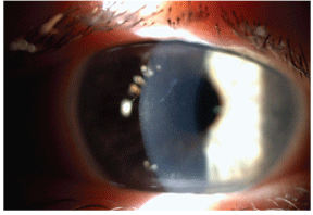
 Epithelial basement membrane dystrophy (EBMD), also known as Cogans microcystic epithelial dystrophy or map-dot-fingerprint dystrophy, is one of the most common anterior segment conditions an eye-care physician will observe in clinical practice. Patients with EBMD often present with severe chronic recurrent corneal erosion (RCE), glare and photophobia; however, some EBMD patients may appear entirely asymptomatic.
EBMD affects nearly 42% of individuals across all age groups, and as many as 76% of individuals worldwide who are over the age of 50.1 Up to 33% of patients with EBMD experience severe RCE during their lifetime.2,3 In an effort to provide information on how to effectively manage this disease, we will review the causes, symptoms and treatment options of EBMD.
EBMD
The clinical signs of epithelial basement membrane dystrophy typically include a bilateral presentation of epithelial microcysts and whirling superficial defects, such as corneal ridges and opacities.
EBMD is not considered an inherited condition, but there are several reports of families that demonstrate an autosomal dominant inherited pattern. In one study, researchers were able to identify two different point mutations in the genes associated with other corneal dystrophies.4

This patient with epithelial basement membrane dystrophy (EBMD) demonstrates map lines. Maps are sharply demarcated areas with hazy, white centers.
EBMD is associated with a faulty basement membrane, which is thickened, multilaminar or redundant, and misdirected into the epithelium. The basal epithelial cells manufacture either unconventional redundancies or finger-like projections that protrude from an abnormally thickened basement membrane.5
Diagnosis
Sometimes, a diagnosis of EBMD can be made with a proper patient history. Dry eye is a common symptom of EBMD, and patients may report fluctuating vision, grittiness or photophobia. In cases associated with RCE, patients may experience a sharp pain upon waking.
In other cases, the diagnosis can be made with keratometry, since the mires will appear irregular. Also, a slit lamp examination can reveal the presence of microcysts, which are epithelial cells trapped in intercellular debris in the redundant basement membrane formation. These microcysts can be observed best with fluorescein staining, and often appear as inverse or negative staining areas. Additionally, corneal imaging is valuable in the diagnosis of EBMD. A slit lamp examination should reveal the classic identifying projections and redundant basement membrane formations.

Recurrent corneal erosion in an EBMD patient.
One interesting finding: Almost 90% of RCE cases, especially those associated with EBMD, occur in the inferior third of the cornea.3,6 So, carefully examine the inferior region of the cornea for breakdown and staining in EBMD patients.
Treatment
In mildly asymptomatic cases, lubrication with artificial tears and/or punctal plugs may be warranted to prevent irritation. In moderate cases, a hypertonic drop or ointment is recommended. One new treatment option is FreshKote (Focus Laboratories), which appears to be effective for EBMD patients with or without associated RCE. FreshKote drops are available by prescription only. The drops work on an oncotic pressure basis, which is similar to hypertonic drops, without adding further hypertonicity upon instillation. A sodium ointment applied at night, such as Muro 128 5% (Bausch & Lomb), may also benefit these patients.
In more advanced cases of RCE associated with EBMD, steroids, such as loteprednol, cyclosporine or low-dose oral doxycycline, such as Alodox (20mg doxycycline hyclate, OCuSOFT), should be considered.7 Previous research has shown that corticosteroids and oral doxycycline are very effective treatments for RCE.8 The mechanism of action occurs through suppression of the enzyme matrix metalloproteinase-9 (MMP-9), which is widely responsible for catalyzing corneal erosion.
Patients treated with a combination of these medications demonstrated rapid resolution and experienced no recurrences of corneal erosion.8 If RCE in EBMD patients is resistant to topical or oral therapy, surgery, such as superficial keratectomy or phototherapeutic keratectomy (PTK), is recommended.9 Both diamond burr polishing of Bowmans layer and PTK were found to be very effective treatments, particularly for EBMD patients who experience recalcitrant RCE.10
In one study of patients with chronic recalcitrant recurrent corneal erosion, 86% experienced successful resolution with photothrerapeutic keratectomy after 12 months.11 Research has demonstrated that patients with EBMD should not be considered as candidates for LASIK. Instead, EBMD patients should undergo PRK or LASEK.12
Patients with EBMD, or even sub-clinical EBMD, can experience severe corneal epithelial sloughing and multiple complications during a LASIK procedure.13 In one such study, 100% of the patients with epithelial sloughing were diagnosed with EBMD either before or after LASIK. Related complications included a high prevalence of epithelial ingrowth (73%), diffuse lamellar keratitis (55%), flap microfolds (18%) and flap melting (36%).14
Epithelial basement membrane dystrophy is one of the most common conditions diagnosed by eye-care professionals. We now have a better understanding of the causes and symptoms of EBMD.
Most importantly, new research is uncovering modern diagnostic methods and treatment options to help our patients combat a serious condition that can rapidly progress and cause severe ocular complications.
Dr. Karpecki is a consultant to Allergan, Bausch & Lomb and OCuSOFT. He has no financial interest in any of the products mentioned.
1. Werblin TP, Hirst LW, Stark WJ, et al. Prevalence of map-dot-fingerprint changes in the cornea. Br J Ophthalmol 1981 Jun;65(6):401-9.
2. Ghosh M, McCulloch C. Recurrent corneal erosion, microcystic epithelial dystrophy, map configurations and fingerprint lines in the cornea. Can J Ophthalmol 1986 Oct;21(6):246-52.
3. Reidy JJ, Paulus MP, Gona S. Recurrent erosions of the cornea: epidemiology and treatment. Cornea 2000 Nov;19(6):767-71.
4. Boutboul S, Black GC, Moore JE, et al. A subset of patients with epithelial basement membrane corneal dystrophy have mutations in TGFBI/BIGH3. Hum Mutat 2006 Jun;27(6):533-7.
5. Labb A, Nicola RD, Dupas B, et al. Epithelial basement membrane dystrophy: evaluation with the HRT II Rostock Cornea Module. Ophthalmology 2006 Aug;113(8):1301-8.
6. Hykin PG, Foss AE, Pavesio C, Dart JK. The natural history and management of recurrent corneal erosion: a prospective randomized trial. Eye 1994;8 (Pt 1):35-40.
7. Sobrin L, Liu Z,
8. Dursun D, Kim MC, Solomon A, et al. Treatment of recalcitrant recurrent corneal erosions with inhibitors of matrix metalloproteinase-9, doxycycline and corticosteroids. Am J Ophthalmol 2001 Jul;132(1):8-13.
9. Itty S, Hamilton SS, Baratz KH, et al. Outcomes of epithelial debridement for anterior basement membrane dystrophy. Am J Ophthalmol 2007 Aug;144(2):288-9.
10. Sridhar MS, Rapuano CJ, Cosar CB, et al. PTK versus diamond burr polishing of Bowmans membrane in the treatment of recurrent corneal erosions associated with anterior basement membrane dystrophy. Ophthalmology 2002 Apr;109(4):674-9.
11. Cavanaugh TB, Lind DM, Cutarelli PE, et al. Phototherapeutic keratectomy for recurrent erosion syndrome in anterior basement membrane dystrophy. Ophthalmology 1999 May;106(5):971-6.
12. Dastjerdi MH, Soong HK. LASEK (laser subepithelial keratomileusis). Curr Opin Ophthalmol 2002 Aug;13(4):261-3.
13. Rezende RA, Uchoa UC, Cohen EJ, et al. Complications associated with anterior basement membrane dystrophy after LASIK. J Cataract Refract Surg 2004 Nov;30(11):2328-31.
14. Perez-Sntonaja JJ, Galal A, Cardona C, et al. Severe corneal epithelial sloughing during LASIK as a presenting sign for silent epithelial basement membrane dystrophy. J Cataract Refract Surg 2005 Oct;31(10) 1932-7.

