
|

|
The most common indications for penetrating keratoplasty are regrafting, keratoconus, Fuchs’ endothelial dystrophy, pseudophakic bullous keratopathy, perforated corneas, traumatic scars and viral keratitis.
The advantages of penetrating keratoplasty include the full removal of damaged corneal tissue, improved optical clarity, restored corneal anatomy, ease of performance compared to other corneal transplant procedures, improved cosmetic appearance and the potential for good optical results.
Some disadvantages are a higher risk of graft rejection, post-op astigmatism, suture management, intraocular complications and traumatic corneal rupture.
Variations of the procedure include deep anterior lamellar keratoplasty (DALK), Descemet’s stripping endothelial keratoplasty (DSEK) and Descemet’s membrane endothelial keratoplasty (DMEK). The choice of procedure (PK or one of the above variations) depends on which corneal layers have been affected.
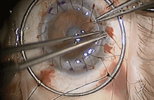
The procedure begins with the preparation of the donor tissue. A trephine (a circular cutting device) is used to cut the donor cornea, followed by trephination of a similar sized portion (7mm to 9mm) of the patient’s cornea. Once the recipient’s corneal button has been removed, the anterior chamber is filled with balanced salt solution or sodium hyaluronate and the donor button is placed into position.
Multimedia
Click
here to view a narrated video of penetrating keratoplasty.
Photo and video courtesy of corneal specialist John Sheppard, MD, MMSc, Virginia Eye Consultants.
Four cardinal sutures of 10-0 nylon are placed at 90˚ intervals in the donor graft, just above Descemet’s membrane. The sutures are then passed into the recipient’s cornea at the same level, at approximately 1.5mm into the host tissue. Once the needle is passed though, the suture is tied and buried.
After the cardinal sutures are in place, suturing can be completed with a single running suture or interrupted sutures.
Postoperatively, patients are prescribed topical antibiotics for one to two weeks as well as topical steroids, which are tapered over several months. Many times, patients are kept on low-dose topical steroids to reduce the risk of graft rejection and failure. Sutures can be removed as soon as two to three months, if needed. Or, if a patient has little astigmatism and the sutures are not causing any problems, they can be left in place for many years.
As comanaging optometrists, our main concern is the long term management and visual function. Postoperatively, corneas may take anywhere from 18 to 24 months to fully stabilize, so be sure to continuously monitor patients for adequate visual acuity and functional vision. Communicate with your corneal specialist to decide when patients are sufficiently stable for contact lenses. A specialty contact lens (GP or hybrid) may be considered as soon as three months after surgery, but may need several changes and modifications once the sutures are removed.
