 A 75-year-old white male presented as a new patient in January 2009. He was recently diagnosed with glaucoma by his previous eye-care provider and was being medicated with one drop Travatan (travoprost, Alcon) h.s. O.U. as well as 0.5% timolol every morning.
He underwent uncomplicated bilateral cataract surgery in 2005. The patients history also suggested that he was monitored for suspicious optic nerves for several years before beginning therapy in the summer of 2008.
His systemic medications included lisinopril for hypertension. The patient reported no known drug allergies.
Diagnostic Data
Best-corrected visual acuity measured 20/25- O.D. and 20/20- O.S. through hyperopic, astigmatic and presbyopic correction. His pupils were equally round and reactive to light and accommodation, with no afferent pupillary defect. Extraocular muscles were full in all positions of gaze.
On initial presentation, a slit lamp examination of his anterior segments appeared unremarkable. Mild corneal arcus and scattered guttatae were present O.U. Intraocular pressure measured 12mm Hg O.D. and 13mm Hg O.S. Pachymetry readings were 526m O.D. and 507m O.S.
Through dilated pupils, his posterior chamber intraocular lenses were well centered in the capsular bags, and the posterior capsules were essentially clear O.U. His optic nerves measured 0.40 x 0.50 O.D. and 0.55 x 0.70 O.S. His inferior temporal retinal rims were thinner O.S. than O.D. Both maculae were characterized by retinal pigment epithelium (RPE) granulation and drusen that centered in the foveal avascular zone (FAZ), O.D. greater than O.S. The maculopathy was consistent with his best-corrected visual acuities. There was a small epiretinal membrane (ERM) located outside of the FAZ O.S.
His vascular examination was unremarkable. His peripheral retinal exam was consistent with 360 of pigmented microcystoid O.U., with scattered areas of pavingstone. There were no holes or tears O.U.; however, the patient demonstrated bilateral posterior vitreous detachments.
I asked the patient to continue his current ophthalmic medication regimen and return in one month for further evaluation. Also, I requested a copy of his previous eye care records.
At one month, we performed Heidelberg Retina Tomography-3 (HRT-3), optic nerve imaging, gonioscopy, threshold white-on-white (WOW) perimetry and tomometry. At this visit, intraocular pressure measured 11mm Hg O.U. Gonioscopy demonstrated IV+ open angles, with grade I trabecular pigmentation O.U. No iris or angle abnormalities were present.
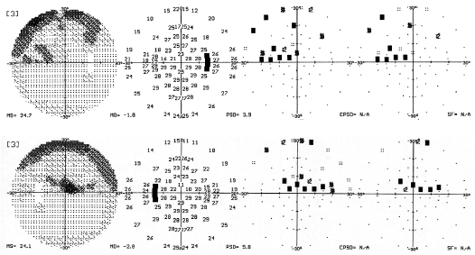
HRT-3 imaging confirmed the asymmetrically thin neuroretinal rims, and Moorfields regression analysis flagged several sectors of each optic nerve as abnormalmost notably the inferior temporal aspects of each rim. Threshold WOW perimetry demonstrated a superior arcuate defect O.D. with nasal step, and a denser superior arcuate defect O.S. that involved fixation (figures 1 and 2).
Given the current clinical picture, I established his target IOP at 12mm Hg to 14mm Hg O.U. Because the patient was nearly at target IOP, I asked him to continue his current medications and return for follow-up in three months for a standard glaucoma visit in conjunction with a flicker-defined form visual field testing.

1, 2. Standard white-on-white (WOW) perimetry results of our patient (O.D. top, O.S. bottom). Note the amount of visual field loss.
Discussion
Evaluation of visual fields is one of the most frustrating aspects of glaucoma management from the perspective of both the patient and the clinician. Visual field studies often yield varied results that are difficult to reproduce because of patient fatigue and other associated subjective factors.
Conventional WOW perimetry has been the gold standard in managing glaucoma patients for many years. However, there are many limitations to standard WOW perimetry, particularly during the early stages of glaucoma, when fewer axons are damaged.
In WOW perimetry, all ganglion cells are believed to be stimulated simultaneously. So, when damage exists across all cell types, visual field defects will be defined easily. However, the earliest damage to ganglion cells occurs preferentially in the M-cell (magnocellular) population.1,2 Therefore, stimuli that specifically target M-cells can uncover damage to the ganglion cell pathway significantly earlier than WOW perimetry.3 By taking advantage of this reduced redundancy process, commercial perimeters, such as the Humphrey Matrix (Carl Zeiss Meditec), have been developed to exploit early susceptibility of the M-cell pathway and highlight field defects earlier.
But, when you consider test/re-test variability, it is clear that many perimeters lack strong re-test reproducibility. And ultimately, reproducible data is the crux of periodic visual field testing.
In standard WOW perimetry, there is tight correlation between test/re-test reliability when the patient has good visual functioning. Consistent reproducibility declines significantly, however, when threshold levels fall below 20dBwhen early field defects become apparent.
The Heidelberg Edge Perimeter (HEP, Heidelberg Engineering) has shown significantly less test/re-test variability across all threshold levels. Furthermore, the HEP uses a flicker-defined form of stimulus that selectively stimulates M-cells.4 So, the HEP may be able to detect visual field defects even earlier than conventional WOW perimetry.

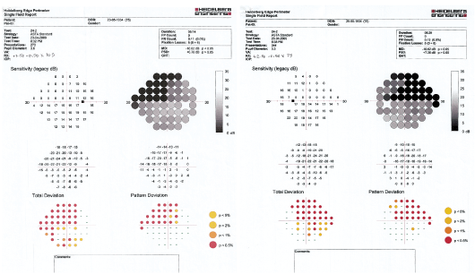
The HEP offers various threshold examinations, including 24-2, 10-2, neurological and custom maps. The HEP printout includes the raw threshold dB sensitivity, a grayscale plot, total deviation and pattern deviation.
Additionally, the visual field printout illustrates statistically significant deviation with a color-coded legend, which ranges from 5% to 0.5% confidence levels.
If our patients former eye-care provider would have performed HEP testing initially, a diagnosis of glaucoma may have been confirmed earlier (figures 3 and 4). In any case, HEP testing will play a significant role in our patients long-term management because we will be able to follow his visual field changes reliably over time.
Dr. Fanelli has no financial interests in any of the technologies mentioned.
1. Glovinsky Y, Quigley HA, Dunkelberger GR. Retinal ganglion cell loss is size dependent in experimental glaucoma. Invest Ophthalmol Vis Sci 1991 Mar;32(3):484-91.
2. Kerrigan-Baumrind LA, Quigley HA, Pease ME, et al. Number of ganglion cells in glaucoma eyes compared with threshold visual field tests in the same persons. Invest Ophthalmol Vis Sci 2000 Mar;41(3):741-8.
3. Racette L, Medeiros FA, Zangwill LM, et al. Diagnostic accuracy of the Matrix 24-2 and original N-30 frequency-doubling technology tests compared with standard automated perimetry. Invest Ophthalmol Vis Sci 2008 Mar;49(3):954-60.
4. Johnson CA. Psychophysical measurement of glaucomatous damage. Surv Ophthalmol 2001 May;45 Suppl 3:S313-8.

