 |
Q:
A concerned patient presented with the chief complaint of a growth under his upper lid. What is my differential and what do I do about it?
A:
“Subconjunctival orbital fat prolapse is a rarely reported, benign condition,” says Paige Thompson, OD, of SouthEast Eye Specialists in Chattanooga, TN. “Most patients present for evaluation due to cosmetic concerns or to rule out potential for malignancy.”
Orbital fat occupies the space surrounding the extraocular muscles and the globe—a protective cushion for the eye. The orbital fat is comprised of two compartments: intraconal and extraconal. Orbital fat may be prolapsed or displaced secondary to aging, trauma or surgery.1,2 With extraconal fat prolapse, the orbital fat migrates beyond the orbital septum and results in lower lid festoons.2 When intraconal fat is displaced through Tenon’s capsule, it can be visualized in the subconjunctival space.1
Subconjunctival orbital fat is most commonly seen in elderly males as a result of age-related weakening of Tenon’s capsule.2,3 This finding may be unilateral or bilateral, but in bilateral cases it is often asymmetric. Subconjunctival herniated orbital fat presents as a soft, mobile, yellow mass with superficial vasculature.1-3 Dr. Thompson says this mass is most often seen in the superior temporal quadrant and can be indented with a cotton tip applicator. Prolapsed orbital fat will become more prominent with pressure applied to the globe or with downward movement of the eye.1 Patients with subconjunctival herniated orbital fat are typically asymptomatic; however, patients may report mild irritation and tearing.4
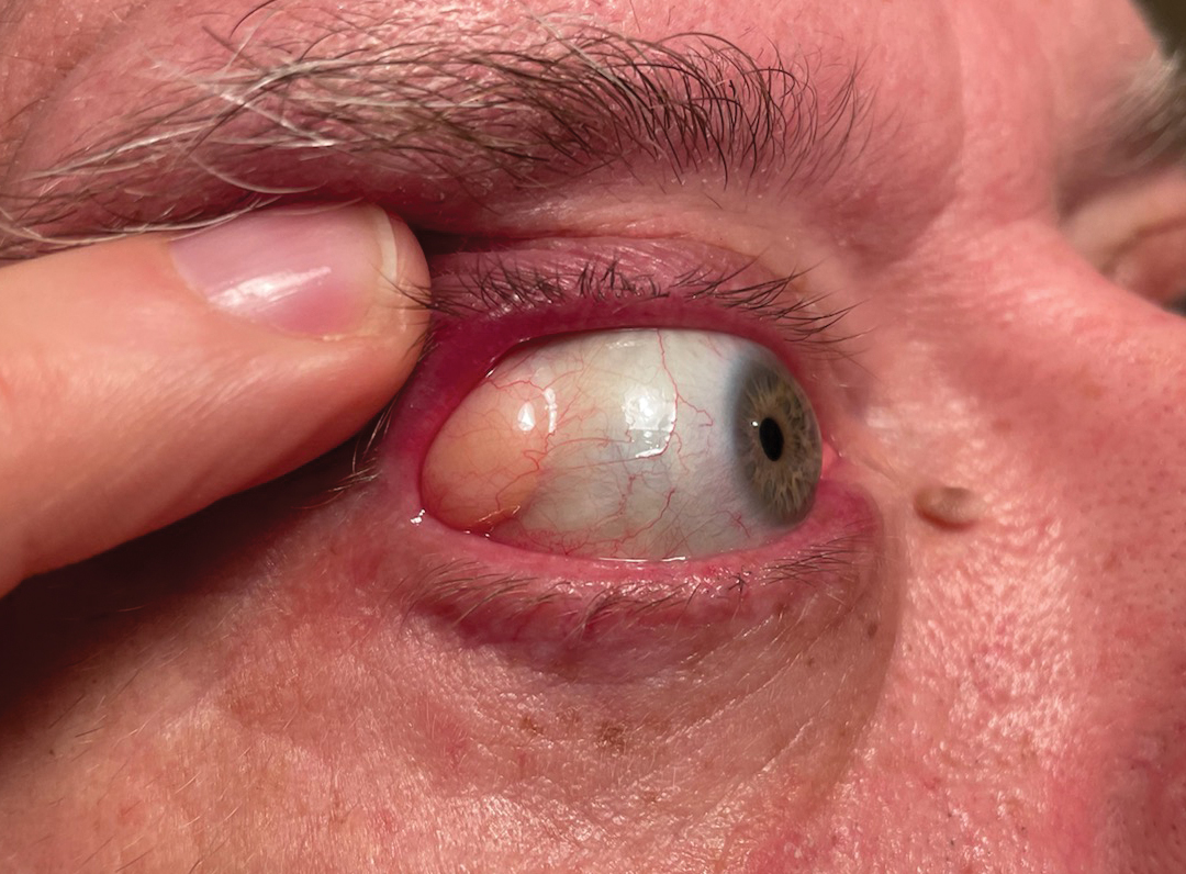 |
| Subconjunctival orbital fat may be prolapsed secondary to aging, trauma or surgery. Click image to enlarge. |
Differential and Treatment
Dermolipoma, lacrimal gland prolapse, lipomatous tumors and lymphoma are all potential differential diagnoses in these cases. A dermolipoma is a congenital lesion of the orbit which presents as an immobile, pinkish-white mass in the superior temporal quadrant of the orbit. Lacrimal gland prolapse is often difficult to visualize without lid eversion. Lipomatous tumors are very rare and can be benign or malignant. These appear as a yellowish-pink mass on the conjunctiva in adults and often require histopathology for definitive diagnosis.
Conjunctival lymphoma presents as a salmon-colored growth that is diffuse and non-mobile. Lymphoma typically grows more rapidly than prolapsed orbital fat, may present with feeder vessels and is firm to the touch.2-4
Most often, subconjunctival herniated orbital fat can be differentiated from other orbital tumors on clinical exam. But in difficult cases, Dr. Thompson suggests that histopathology may be required to make a definitive diagnosis.
“Neuroimaging with MRI or CT can also clearly demonstrate prolapsed orbital fat, but are indicated only when a diagnosis cannot be made based on clinical exam,” she says. On neuroimaging, subconjunctival orbital fat prolapse is continuous with the intraconal fat, unlike many other orbital lesions.5
Treatment of subconjunctival orbital fat prolapse typically involves observation. In some cases, a patient may elect to have the mass removed due to symptomatology, cosmetic concerns or suspicion for malignancy. Most often, prolapsed fat is accessed through a transconjunctival incision. Orbital fat is typically resected and the incision is then closed with sutures or fibrin glue.2,4,6 This procedure is performed on an outpatient basis and involves local anesthesia.3 Some surgeons prefer to reposition the orbital fat rather than resect it; however, resection is still the most common approach. Risks of surgical resection include infection, lacrimal gland damage and retrobulbar hemorrhage.4
“Fortunately, this procedure has a high success rate and risk of recurrence is low,” Dr. Thompson says.
Dr. Ajamian is the center director of Omni Eye Services of Atlanta. He currently serves as general chairman of the education committee for SECO International. He has no financial interests to disclose.1. Schmack I, Patel RM, Folpe AL, et al. Subconjunctival herniated orbital fat: a benign adipocytic lesion that may mimic pleomorphic lipoma and atypical lipomatous tumor. Am J Surg Pathol. 2007;31(2):193-8. 2. Skorin L. Subconjunctival orbital fat prolapse: Diagnosis and management. Optometric Clinical Practice. 2019;1(1). 3. Khalil D, King B. A unilateral orbital mass. JAMA Dermatol. 2014;150(10):1115. 4. Secondi R, Sánchez España JC, Castellar Cerpa J, Ibáñez Flores N. Subconjunctival orbital fat prolapse: an update on diagnosis and management. Semin Ophthalmol. 2019;34(2):69-73. 5. Kim E, Kim HJ, Kim YD, Woo KI, Lee H, Kim ST. Subconjunctival fat prolapse and dermolipoma of the orbit: differentiation on CT and MR Imaging. AJNR Am J Neuroradiol. 2010;32(3):465-7. 6. Siban M, Weijtens O, van den Bosch W, Paridaens D. Efficacy of transconjunctival excision of orbital fat prolapse: a long-term follow-up study. Acta Ophthalmol. 2014;92(3):291-3. |
