 In our previous two “Double Trouble” articles (
September and
November 2012), we described two patients being treated for glaucoma who concurrently developed double vision from cranial nerve (CN) VI and CN III palsies, respectively. This month, we complete the trifecta.
In our previous two “Double Trouble” articles (
September and
November 2012), we described two patients being treated for glaucoma who concurrently developed double vision from cranial nerve (CN) VI and CN III palsies, respectively. This month, we complete the trifecta.
A 54-year-old man being treated for primary open-angle glaucoma reported occasionally experiencing double vision. Because the double vision was only intermittent, the patient wasn’t overly concerned––he simply wanted to express the complaint.
Upon questioning, he said that the double vision was vertical and became worse when he was reading and/or tired. He did not report any other physical problems and said that he felt well overall. His recent physical examination was normal, save a slightly elevated cholesterol level.
On general inspection, it was easy to see that he had a slight head tilt to the right. Alternate cover testing demonstrated a left hyperphoric deviation, which worsened in right gaze and left head tilt. Based upon this signature motility, he was diagnosed with a left CN IV palsy.
What is CN IV Palsy?
Patients with CN IV palsy typically present with complaints of vertical diplopia that worsens when reading. There may be an inability to look both down and in. There may also be a component of horizontal diplopia, as a lateral phoria becomes manifest due to the vertical dissociation.1-4 The patient’s chin also may be tucked downward as well.
Further, the patient may report greater diplopia or visual discomfort when tilting his or her head toward the side of the palsy. Commonly, the patient develops a compensatory head tilt opposite to the affected superior oblique muscle. Accomodating for these postural changes generally makes the patient more visually comfortable. Ocular motility testing with the alternate cover test will reveal a hyperphoric or hypertropic deviation that worsens upon opposite gaze and same-side head tilt.5-8
Patients with CN IV palsy frequently present with concurrent hypertension and/or diabetes.9-11 In many instances, there will be a history of head trauma immediately preceding development of CN IV palsy. The trauma need not be major, as relatively minor injuries can trigger the event.2,3,12-14 In cases of longstanding, decompensated CN IV palsy, the inciting trauma may have occurred several years earlier and often is forgotten by the patient.
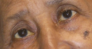
Another patient with a congenital left CN IV palsy and a severe right head tilt.
The fourth nerve especially is prone to trauma, because it exits the brain stem and courses through the subarachnoid space. In contrast to third nerve palsies with an etiology in the subarachnoid space, fourth nerve palsies are rarely caused by aneurysmal compression. The most common causes of damage to the fourth nerve in this region are trauma and ischemic vasculopathy.3
Due to the large number of other neural structures that accompany the fourth nerve as it travels through the cavernous sinus and superior orbital fissure, it is unlikely that patients will exhibit isolated fourth nerve palsy due to damage within these areas. More likely, there will be a concomitant palsy of cranial nerves III and VI. Common causes of damage to the fourth nerve in these areas are herpes zoster, inflammation of the cavernous sinus or posterior orbit, meningioma, metastatic disease, pituitary adenoma and carotid cavernous fistula.15 Trauma to the head or orbit can cause damage to the trochlea with resultant superior oblique muscle dysfunction.
Trauma and vascular disease are considered the main causes of acquired CN IV palsy.14,15 However, numerous reports of other potential causes of isolated CN IV palsy, include multiple sclerosis, polycythema vera, cat-scratch disease and, rarely, metastatic
disease.15
Managing Patients with CN IV Palsy
A fourth nerve palsy often presents suddenly, but may result from decompensation of a longstanding or congenital palsy and the onset just seems sudden. In order to differentiate these two types of palsies, ask for old photographs. A patient with a decompensated, longstanding palsy will present with a compensatory head tilt that often can be identified in photographs. Usually, patients are not even aware of their head tilt.
Patients with longstanding, decompensated fourth nerve palsies will have an exaggerated vertical fusional ability. Such longstanding fourth nerve palsies typically have a benign course that requires no further management.
In the case of complicated fourth nerve palsies (those that present with other concurrent neurological dysfunction), the patient should undergo neuroradiological studies. The accompanying signs and symptoms typically dictate the extent of these evaluations.
In the case of isolated fourth nerve palsies caused by recent trauma, the patient should also undergo neuroradiological studies of the head to dismiss the possibility of a concurrent subarachnoid hemorrhage. If the fourth nerve palsy is not associated with recent trauma, a history of past trauma should be investigated.
If the fourth nerve palsy is due to previous trauma and has recently decompensated, the diplopia can be managed by the placement of vertical prisms in spectacles. Further, if the patient is elderly and has a fourth nerve palsy of truly recent origin, an ischemic vascular evaluation should be undertaken to search for diabetes and hypertension.
If, however, the palsy is caused by vascular infarct, then it will spontaneously resolve over a period of three to six months. Usually, no further management beyond periodic observation and either occlusion or press-on prism therapy is required.2,3 But, in some cases, recovery does not occur.2,3 In these instances, permanent prism (ground into the spectacle lenses), muscle surgery or botulinum toxin A injections may be considered.16
When encountering isolated CN IV palsy, delay prescribing permanent prisms for at least three months in order to allow for the palsy to recover. Otherwise, glasses with permanent prism correction can induce vertical diplopia, should the palsy recover.
In the case presented here, the patient was educated about his new diagnosis. His recent normal physical exam and lack of other signs or symptoms, along with the head tilt, made us suspect that the patient had a longstanding CN IV palsy that had simply decompensated. We confirmed our suspicions when he produced his driver’s license photo (which was several years old), showing the right head tilt.
A 1.00∆ base-down prism over his left spectacle lens made him feel more visually comfortable. Going forward, we instructed him to remain keenly aware of his double vision. Additionally, we informed him that if the problem persisted and affected his quality of life, then we’d add the prismatic correction to his spectacles permanently.
1. Staubach F, Lagrèze WA. Oculomotor, trochlear, and abdu- cens nerve palsies. Ophthalmologe. 2007 Aug;104(8):733-46.
2. Akagi T, Miyamoto K, Kashii S, et al. Cause and prognosis of neurologically isolated third, fourth, or sixth cranial nerve dysfunction in cases of oculomotor palsy. Jpn J Ophthalmol. 2008 Jan-Feb;52(1):32-5.
3. Hoya K, Kirino T. Traumatic trochlear nerve palsy following minor occipital impact––four case reports. Neurol Med Chir (Tokyo). 2000 Jul;40(7):358-60.
4. von Noorden GK, Murray E, Wong SY. Superior oblique paralysis. A review of 270 cases. Arch Ophthalmol. 1986 Dec;104(12):1771-6.
5. Baumeister E. Contribution to the diagnosis of trochlear paresis (first description of Bielschowsky head-tilt test). 1874. Strabismus. 2003 Jun;11(2):129-30.
6. Simonsz HJ, Crone RA. Bielschowsky head-tilt test––I. Ocular counterrolling and Bielschowsky head-tilt test in 23 cases of superior oblique palsy. Vision Res. 1985;25(12):1977-82.
7. Gräf M, Krzizok T, Kaufmann H. Head-tilt test in unilateral and symmetric bilateral acquired trochlear nerve palsy. Klin Monbl Augenheilkd. 2005 Feb;222(2):142-9.
8. Straumann D, Bockisch CJ, Weber KP. Dynamic aspects of trochlear nerve palsy. Prog Brain Res. 2008;171:53-8.
9. Park UC, Kim SJ, Hwang JM, Yu YS. Clinical features and natural history of acquired third, fourth, and sixth cranial nerve palsy Eye. Eye (Lond). 2008 May;22(5):691-6.
10. Trigler L. Retinopathy in patients with diabetic ophthalmoplegia. Ophthalmology. 2003 Aug;110(8):1545-50.
11. Acaroglu G. Retinopathy in patients with diabetic ophthalmoplegia. Ophthalmologica. 2008;222(4):225-8.
12. Dhaliwal A, West AL, Trobe JD, Musch DC. Third, fourth, and sixth cranial nerve palsies following closed head injury. J Neuroophthalmol. 2006 Mar;26(1):4-10.
13. Ishizaki E, Kurokawa Y. A case of solitary and unilateral trochlear nerve palsy due to a blunt head impact. Rinsho Shinkeigaku. 2003 Sep;43(9):571-3.
14. de Camargo GB, Hida WT, Goldchmit M, et al. Paralytic strabismus: review of 24 years at “Santa Casa de São Paulo.” Arq Bras Oftalmol. 2007 Jul-Aug;70(4):585-7.
15. Richards BW, Jones FR Jr, Younge BR. Causes and prognosis in 4,278 cases of paralysis of the oculomotor, trochlear, and abducens cranial nerves. Am J Ophthalmol. 1992 May;113(5):489-96.
16. Bagheri A, Eshaghi M. Botulinum toxin injection of the inferior oblique muscle for the treatment of superior oblique muscle palsy. J AAPOS. 2006 Oct;10(5):385-8.