 |
|
|
In
jected steroid use has become more
common for ODs
.
|
The mainstay treatment for ocular inflammation is corticosteroid therapy, and every practicing optometrist knows the inherent risk of increasing a patient’s intraocular pressure (IOP) with the addition of a steroid. We stress this fact so often that young practitioners are apprehensive about using corticosteroids effectively. When prescribed responsibly, corticosteroids continue to be a valuable component of our pharmaceutical armamentarium against inflammation in our glaucoma patients.
Basic Pharmacology of Corticosteroids
Inflammation involves the activation and proliferation of many types of chemical messengers and immune cells, including cytokines, macrophages and prostaglandins.1 Since the 1950s, corticosteroids—or just “steroids” for most of us—have been shown to effectively treat most types of inflammation.1 Steroids’ anti-inflammatory effect is due to their ability to directly or indirectly alter gene transcription. Corticosteroids penetrate cell membranes and bind to receptors in the cytoplasm, yielding a conformational change in the receptor and facilitating transportation of the receptor-steroid complex into the cell nucleus, where it binds to specific sequences on DNA.2 Genes activated by corticosteroids include those that decrease inflammatory signal transduction, inhibit macrophage function and diminish the distribution of leukocytes.1,2 Another effect of corticosteroids is their ability to deactivate genes that code for synthesis of prostaglandins, leukotrienes and platelet-activating factor. Additionally, they reduce the expression of cyclooxygenase-2, which leads to a further decrease in prostaglandin synthesis.2 It has been shown that corticosteroids render an additional effect at the level of mRNA, leading to further reduction in protein synthesis.1It is important to note that widespread use of corticosteroids can cause multiple adverse reactions. Tissue and metabolic changes, such as facial swelling, fat redistribution, increased hair growth, weight gain and muscle weakness, have all been shown in patients on long-term corticosteroid treatment. Other changes include hyperglycemia, glucose intolerance, high blood pressure and osteoporosis. Of greater interest to optometrists, however, is the potential for posterior subcapsular cataracts and increased IOP.3
Mechanism of Effect on IOP
The primary association between corticosteroid use and elevated IOP seems to be decreased aqueous outflow secondary to the aggregation of extracellular matrix material in the trabecular meshwork (TM).4 More specifically, one study indicated that corticosteroids reduce the release of chemicals responsible for mucopolysaccharide degradation in the TM.5 Accumulation of these mucopolysaccharides likely is responsible for increased aqueous outflow resistance. It has also been shown that corticosteroids may cause a reversible crosslinking of actin fibers within TM cells, further contributing to increased outflow resistance.6 Several studies have compared the steroid response of patients both with and without glaucoma.7-10 J.Francois, MD, published the first case report on corticosteroid-induced glaucoma in 1954.7 He suggested that IOP increase occurs within six to 12 months in patients on mild corticosteroids (e.g., prednisone), but could take just a few weeks for patients on more potent agents (e.g., dexamethasone).7 Then in 1963, Bernard Becker, MD, and Donald Mills, MD, showed that patients who were previously diagnosed with open-angle glaucoma or were identified as glaucoma suspects exhibited a much greater IOP response to corticosteroids than healthy controls.8In 1975, P.F. Palmberg, MD, PhD, and associates documented that IOP increase secondary to corticosteroid dosing was consistent and repeatable on the same patient over different periods of time.9 Ten years later, Robert N. Weinreb, MD, and associates showed that the IOP response is significantly more rapid in patients with previously diagnosed glaucoma than in those with a normal IOP.10 It is worth noting that both the Becker and Mills study and the Weinreb study showed that corticosteroid response in glaucoma patients occurred independently of the patient’s treatment status. Considering these study data, it appears that glaucoma patients on corticosteroid thrapy are much more likely to experience an IOP increase than the rest of our patients. Thus, we must remain extremely cautious when choosing an anti-inflammatory therapy for these individuals.
Steroid Use in Eye Care
• Topical corticosteroids are commonly used to treat a host of ophthalmic conditions, including allergic reactions of the eyelids, conjunctiva or cornea; scleritis and episcleritis; anterior and posterior uveitis; giant-cell arteritis; scar prevention following ocular trauma; herpes zoster ophthalmicus; and practically any other ocular condition that involves inflammation.11Topical dosing delivers therapeutic drug levels to the cornea and aqueous humor. Numerous forms of conjunctivitis, as well as episcleritis, scleritis, anterior uveitis and other anterior segment diseases, respond well to topical therapy.12 Another study by Dr. Becker showed that topical corticosteroids produced an IOP response similar to that associated their systemic counterparts, and that the response also was greater in patients with glaucoma than in those with normal IOP measurements.12 If topical eyelid or adnexa treatment is required, dermatological preparations or ocular ointments also can elevate IOP.13
Further, pressure increases have been shown in patients who use inhaled or nasal steroids.14
• Systemic steroid therapy is very common, and many of our patients will use these agents for one reason or another. In optometric practice, it’s sometimes necessary to add a systemic corticosteroid when posterior segment inflammation is involved or if anterior segment inflammation is not responding to topical therapy. It should be no surprise that treatment with systemic corticosteroids increases IOP in some patients; however, it is somewhat unusual that the response often is less significant—or takes longer to manifest—than that seen in patients on topical therapy.15 This consideration is important to remember when scheduling follow-up visits.
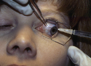
• Injected steroid use has become more common for optometrists, as scope of practice laws have been updated and expanded in certain states. Subconjunctival preparations of corticosteroids have been made available for use in patients who do not respond well to topical treatments or those who are unable to apply topical medications (i.e., severe arthritis). Intraocular pressure response to injected steroids typically is lengthier and more pronounced than that caused by topical corticosteroid use.16 While a topical medication can be discontinued rapidly, an injection cannot be reversed, and natural IOP lowering will not occur until the medication has completely dissipated.16 Intravitreal corticosteroid injections also produce an effect on IOP, but the increase often is delayed beyond the point that we might expect the reaction to occur.17
Corticosteroid Use in Glaucoma Patients
We’ve extensively discussed the greater risk of steroid-induced IOP elevation in our glaucoma patients. So how, then, are we to manage external or internal ocular inflammation in this population? The key is to make responsible decisions in corticosteroid selection and then follow the patient diligently so that consequent IOP increase can managed properly. Corticosteroids differ in their ability to produce an IOP response. In general, the more potent the drug, the greater the hypertensive effect.4 Dexamethasone has the greatest potential to increase IOP, followed by prednisolone, fluoromethalone and hydrocortisone.18 The typical timeframe for a patient to exhibit an IOP with these medications is three to six weeks.18Difluprednate is a relatively new topical corticosteroid that shows increased penetration into the eye and increased bioavailability. Unfortunately, it has also been shown to produce a greater IOP response over a shorter period when compared to prednisolone.19Loteprednol was developed with a different chemistry than other drugs in this class. The structural replacement of a ketone with an ester makes it possible for loteprednol to be metabolized by esterases—thus limiting the potential side effects of this medication.20 One study showed significant decreases in ocular hypertensive effects with loteprednol, without severe reductions in anti-inflammatory activity.20
It’s advisable to avoid corticosteroids in patients with glaucoma—but that’s not always possible.
When a corticosteroid is needed, it’s better to use the least potent agent at the smallest possible dose that still yields a desirable anti-inflammatory effect.4 Typically, my first choice is either loteprednol or fluoromethalone. Then, if neither agent proves effective, I will switch to prednisolone or difluprednate—but only in doses small enough to produce a therapeutic effect.While your patient is on a corticosteroid, it’s important to monitor his or her IOP more closely than normal. A baseline measurement should be taken before therapy is initiated, as well as two to three weeks after. IOP should then be measured every three to four weeks while the corticosteroid therapy is ongoing. If your patient is undergoing intravitreal corticosteroid treatment, IOP should be monitored every two to three weeks for several months following the injection.4
Management of Increased IOP
Corticosteroid-induced IOP increases in the non-glaucomatous population are relatively easy to manage. In most instances, IOP returns to baseline within one to four weeks after treatment discontinuation. The IOP responds to treatment with most of our widely used topical anti-glaucoma medications, including beta-blockers, prostaglandin analogues, alpha agonists, carbonic anhydrase inhibitors and miotics.4 Keep in mind that the management process becomes more complicated when a patient exhibits a steroid response while already on a glaucoma medication. Latanoprost has been shown to be effective in lowering IOP in patients with corticosteroid-induced glaucoma.21 However, it also has been shown to cause anterior segment inflammation, including uveitis. Therefore, the prostaglandin analogues might not be the best first choice to add to a patient who is undergoing treatment for uveitis.4 Further, one study indicated that long-term brimonidine can cause an anterior uveitis after one year or more of continuous dosing.22 This finding should not prevent a clinician from using brimonidine to treat steroid-induced IOP increases altogether. Nevertheless, it’s something to consider as a possible cause of uveitis in a glaucoma patient. Beta blockers and carbonic anhydrase inhibitors are both very effective in controlling corticosteroid-induced glaucoma, and should be considered the “first-line” choice for patients unless otherwise contraindicated.The side effects of corticosteroid use and the risk of increased IOP in glaucoma patients should not deter us from using corticosteroids. When appropriate, the clinician should choose a less potent topical corticosteroid at a smaller dose than usual, and make adjustments based on the patient’s response to the therapy. No matter which medication is selected, IOP must be monitored every few weeks while the patient remains on the medication or for a few months after intravitreal injection. Prudent use, not avoidance, is the key to effective treatment of inflammation in glaucoma patients.
Dr. Ensor is an assistant professor at the Southern College of Optometry in Memphis. He has no industry disclosures or direct financial interest in any of the products mentioned.
1. Barnes PJ. How corticosteroids control inflammation: Quintiles prize lecture 2005. Br J Pharmacol. 2006 Jun;148(3):245-54. 2. Katzung BG, Masters SB, Trevor AJ. Basic & Clinical Pharmacol. 12th ed. New York: McGraw-Hill; 2012. 3. Brenner GM, Stevens CW. Pharmacol. 4th ed. Philadelphia: Elsevier; 2013.4. Kersey JP, Broadway DC. Corticosteroid-induced glaucoma: a review of the literature. Eye (Lond). 2006 Apr;20(4):407-16. 5. Armaly MF. Effect of corticosteroids on intraocular pressure and fluid dynamics: II. The effect of dexamethasone in the glaucomatous eye. Arch Ophthalmol. 1964 May;71:636-44.6. Clark AF, Wilson K, McCartney M, et al. Glucocorticoid-induced formation of cross-linked actin networks in cultured human traecular meshwork cells. Invest Ophthalmol Vis Sci. 1994 Jan;35(1):281-94. 7. Francois J. Corticosteroid glaucoma.Ann Ophthalmol. 1977 Sep;9(9):1075-80.8. Becker B, Mills D. Corticosteroids and intraocular pressure. Arch Ophthalmol. 1963 Oct;70:500-7.9. Palmberg PF, Mandell MD, Wilensky JT, et al. The reproducibility of the intraocular pressure response to dexamethasone. Am J Ophthalmol. 1975 Nov;80(5):844-56.10. Weinreb RN, Polansky JR, Kramer SG, Baxter JD. Acute effects of dexamethasone on intraocular pressure in glaucoma. Invest Ophthalmol Vis Sci. 1985 Feb;26(2):170-5. 11. Dinning WJ. Steroids and the eye – indications and complications. Postgrad Med J. 1976 Oct;52(612):634-8.12. Becker B. Intraocular pressure response to topical corticosteroids. Invest Ophthalmol. 1965 Apr;4:198-205.13. Cubey RB. Glaucoma following the application of corticosteroid to the skin of the eyelids. Br J Dermatol. 1976 Aug;95(2):207-8.14. Garbe E, LeLorier J, Boivin JF, Suissa S. Inhaled and nasal glucocorticoids and the risks of ocular hypertension or open-angle glaucoma. JAMA. 1997 Mar 5;277(9):722-7.15. Bernstein H, Schwartz B. Effects of long-term systemic steroids on ocular pressure and tonographic values. Arch Ophthalmol. 1962 Dec;68:742-53.16. Kalina RE. Increased intraocular pressure following subconjunctival corticosteroid administration. Arch Ophthalmol. 1969 Jun;81(6):788-90.17. Smithen LM, Ober MD, Maranan L, Spaide RF. Intravitreal triamcinolone acetonide and intraocular pressure. Am J Ophthalmol. 2004 Nov;138(5):740-3. 18. Cantrill H, Palmberg P, Zink H, et al. Comparison of in vitro potency of corticosteroids with ability to raise intraocular pressure. Am J Ophthalmol. 1975 Jun;79(6):1012-7.19. Meehan K, Vollmer L, Sowka J. Intraocular pressure elevation from topical difluprednate use. Optometry. 2010 Dec;81(12):658-62. doi: 10.1016/j.optm.2010.09.001.20. Amon M, Busin M. Loteprednol etabonate ophthalmic suspension 0.5%: efficacy and safety for postoperative anti-inflammatory use. Int Ophthalmol. 2012 Oct;32(5):507-17. Epub 2012 Jun 16. 21. Scherer W, Hauber F. Effect of latanoprost on intraocular pressure in steroid-induced glaucoma. J Glaucoma. 2000 Apr;9(2):179-82.22. Byles D, Frith P, Salmon J. Anterior uveitis as a side effect of topical brimonidine. Am J Ophthalmol. 2000 Sep;130(3):287-91.

